
Jon D. Holmes, DMD, MD, FACS
- Assistant Clinical Professor, Department of Oral and
- Maxillofacial Surgery
- University of Alabama at Birmingham
- Private Practice - Oral and Facial Surgery of Alabama
- Birmingham, Alabama
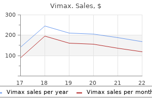
An autopsy revealed a dissection extending from a point 1 to 2 cm proximal to the stent graft to the heart erectile dysfunction statistics uk order discount vimax on-line. Three other subjects expired due to pneumonia erectile dysfunction due to diabetes icd 9 generic vimax 30caps with amex, respiratory failure erectile dysfunction when cheating buy discount vimax on-line, and multisystem organ failure erectile dysfunction depression treatment cheap vimax master card. The subject continues to be active in the trial albeit with permanent adverse sequelae erectile dysfunction in the military purchase vimax with mastercard. Two of the 3 subjects continue to be active in the study erectile dysfunction 45 year old male buy discount vimax online, one with ongoing paraparesis and the other with paraparesis resolved 5 days post surgery. The third subject died 21 days post-procedure due to respiratory failure and had continuing paraparesis at time of death. Each death occurred within the first 30 days and was therefore classified as aneurysm related per protocol. Censored subjects will include those who withdraw, are lost to follow-up, or die from causes adjudicatedto be unrelated to the aneurysm. Kaplan-Meier Estimates of Freedom from Aneurysm-Related Mortality within 12 Months Aneurysm Rupture and Conversion to Surgery No subject experienced aneurysm rupture or conversion to open surgical repair within 12 months. Summary of Primary Effectiveness Endpoint: Valiant Test Group only There were three subjects considered treatment failures in the Valiant Test Group. Two subjects were found to have aneurysm 52 growth of more than 5 mm and had secondary procedures after 365 days (Figure 68. One subject had a distal type Ib endoleak for which a secondary procedure was recommended at the 12-month visit and subsequently performed after 365 days. The other subject had no endoleak per core laboratory at 12 months; a secondary endovascular procedure was performed to relinethe graft when the investigational sitereported continued aneurysm growth without radiographic evidenceof endoleak at the 24-month visit. Change in Maximum Aneurysm Diameter from One Month Secondary Effectiveness Endpoints A summary of secondary effectiveness endpoints is presented in Figure 69. In addition to these secondary endpoints, an evaluation of stent graft integrity was also performed. The other 4 subjectshad no clinical sequelaerelated to endoleak and werealiveat the most recent study visit. No endoleak was reported by theinvestigational sitethough the 24 monthvisit and the subject had no clinical sequelaerelated to endoleak. No lossof stent graft integrity was reported by core laboratory, though the 6 and 12-month x-rayimagescould not beevaluated for stent graft integrity. There was no site-reported lossof stent graft integrity through the 24-monthvisit. Two of the 3 subjectshad limited or no remaining stent graft coverage of the distal nonaneurysmal neck. If thethoracic treatment site cannot be accessed with the delivery catheter, it isconsidered atechnical failure. Two other subjectshadmisaligned deployment, and one subject had an aortic rupture. Subjects diagnosed with a variety of thoracic aortic diseases were considered candidates for the registry. Subjects who enrolled in the study will be followed for up to 3 years post-implantation. A 30-day interim analysis was conducted on 50 subjects to assess acute performance of the Captivia delivery system. Since the acute deliverability of the delivery system is less dependent upon the type of aortic etiology, subjects with dissection and other etiologies were also considered relevant to the assessment. Three subjects died and 1 subject was converted to open surgical repair within 30 days. Thirty-four of the 45 eligible subjects had a follow-up visit at 30 days post-implant. All of the remaining 11 eligible subjects were alive and underwent clinical evaluations at subsequent follow-up visits. Successful delivery and deployment was defined as deployment of the Valiant thoracic stent graft in the planned location with no unintentional coverage of the left subclavian artery, left common carotid artery or brachiocephalic artery, and with the removal of the delivery system. This subject, who had risk factors for neurologic complications, also experienced paraplegia that resolved 2 days later after placement of a lumbar drain. A third death occurred in a subject with a history of Marfans syndrome and previous thoracic aortic dissection. The death was adjudicated as being related to the lesion in an acute complicated type B dissection. One subject required a conversion to open surgery following aneurysm rupture at the index procedure. The subject became unstable after the first stent graft was successfully delivered and deployed. The subject underwent a thoracotomy and a second stent graft was placed, successfully sealing off the rupture site. Two subjects, including the subject with Marfans syndrome noted above, experienced aortic dissection within 30 days of the index procedure. Disease etiologies included fusiform aneurysms and saccular aneurysms/penetrating ulcers of the descending thoracic aorta. A 30-day analysis was conducted on 10 subjects to assess the acute performance of the Captivia delivery system. The data collected from this evaluation was considered relevant because the delivery systems for use with Talent and Valiant stent grafts are essentially identical in design and possess the same principles of operations. Study Population and Subject Accountability these 10 subjects with descending aortic aneurysms were enrolled at 4 sites in the United States to participate in the Talent Captivia Study. Of the 10 enrolled subjects, 1 subject died and another failed to receive a stent graft. Successful Delivery and Deployment Delivery and deployment of the Talent thoracic stent graft with the Captivia delivery system was assessed. Successful delivery and deployment was defined as attaining vessel access to insert the delivery catheter and deployment of the graft to the intended treatment site. One enrolled subject did not receive a Talent thoracic stent graft, as the Captivia delivery system could not reach the targeted lesion due to severe angulation of the thoracic aortic arch. Secondary Study Endpoints Secondary study endpoints evaluated in the 30-day analysis included both procedural complications and clinical outcomes. One subject died within 30 days of the index procedure and was considered an aneurysm related death. Both subjects who experienced paraplegia had significant risk factors for spinal cord ischemia. Caution: Vessel damage such as dissection, perforation, or rupture may be caused by excessive oversizing of the stent graft in relation to the diameter of the blood vessel. Oversizing of the stent graft to the vessel more than the recommended device sizing as shown in Recommended Device Sizing (Section 10. Also, due to the nature of the design and the flexibility of the Valiant thoracic stent graft with the Captivia delivery system, the overall length of each stent graft component may be shorter when deployed. Use of the device outside the recommended anatomical sizing may result in serious device related events. Physicians may consult with a Medtronic representative to determine proper stent graft component dimensions based on the physicians assessment of the patients anatomical measurements. However, the final treatment decision is at the discretion of the physician and patient. The benefits and risks previously described should be carefully considered for each patient before using the Valiant thoracic stent graft with the Captivia delivery system. Medtronic recommends that the physician disclose to the patient, in written form, all risks associated with treatment using the Valiant thoracic stent graft with the Captivia delivery system. The list of potential risks occurring during and after implantation of the device are provided in Adverse Events (Section 5. Additional counseling information can be found in the Patient Information Booklet. Sterility Each Valiant thoracic stent graft is individually contained within a Captivia delivery system. The Captivia delivery system is sterilized using electron beam sterilization and is supplied sterile for single use only. Caution: the Valiant thoracic stent graft with the Captivia delivery system should only be used by physicians and medical personnel trained in vascular interventional techniques and in the use of this device. Recommended Device Sizing Medtronic recommends that the Valiant thoracic stent graft with the Captivia delivery system be used according to the sizing guidelines in Table 3 through Table 8. If preoperative case planning measurements are not certain, an inventory of system lengths and diameters necessary to complete the procedure should be available to the physician. This approach allows for greater intraoperative flexibility to achieve optimal procedural outcomes. Use of the device outside the recommended anatomical sizing may result in serious device related adverse events or clinical incident. The specific stent graft diameter used for treatment should be oversized relative to the nondiseased vessel using the sizing guidelines to ensure appropriate radial fixation. Strict adherence to the sizing guidelines is expected when selecting the appropriate device size. Sizing outside of this range can result in endoleak, fracture, migration, infolding, or graft wear. Caution: Oversizing of the stent graft to the vessel by more than 10% may be unsafe in the presence of dissecting tissue or intramural hematoma. Caution: Proper sizing of the Valiant thoracic stent graft is the responsibility of the physician. This stent graft sizing incorporates the recommended device oversizing for anatomical dimension and was based on in-vitro test data. If it is supported by the vessel, oversizing to the supporting native vessel should be used, as described in Table 3 to Table 6. In order to provide the appropriate oversizing at a component junction that is not supported by the vessel and at the distal landing zones, Closed Web Tapered configurations may need to be used. The order of deployment when using multiple stent graft configurations may vary, depending on the diameter of the aorta proximal to and distal to the lesion. Table 2 should be followed to determine the order of deployment when using multiple stent graft configurations. Caution: When treating acute dissections with multiple devices, it is recommended to deploy the proximal device first. Note: If the vessel diameter and condition require variable proximal and distal diameter configurations, the smallest diameter stent 56 graft should be placed first, either at the proximal or distal end of the lesion. Caution: A FreeFlo or Bare Spring Straight end should never be placed inside the covered section of another stent graft. Order of Deployment When Using Multiple Stent Graft Component Sections Proximal Aortic Diameter = Distal Aortic Proximal Aortic Diameter > Distal Aortic Proximal Aortic Diameter < Diameter Diameter3 Distal First Section Proximal Main Section implanted at Distal Main Section (or other configuration if more Proximal Main Section implantedAortic Diameter Implan ted proximal end of lesion appropri ate) implanted at distal end of lesion at prox imal end of lesion Second Section(Primary Section) Distal Main Section implanted with correct Proximal Main Section implanted with correct Distal Main Section implanted with Implanted junction oversizing. Sizing Guidelines for Treatment of Blunt Traumatic Aortic Injury Native Vessel Suggested Graft Oversizing (mm) (mm) (mm) 18 22 4 Nat Su O ive(m gge(m ve( Vem)19 stem)22 rsim3 sse d mzi 20l Gra22 ng2) 21 22ft 1 22 24 2 23 24 1 24 26 2 25 26 1 26 28 2 27 28 1 28 30 2 29 32 3 30 32 2 31 34 3 32 34 2 33 36 3 34 36 2 35 38 3 36 38 2 37 40 3 38 40 2 39 42 3 40 42 2 40 44 4 41 44 3 42 44 2 42 46 4 43 46 3 44 46 2 10. Dissection Sizing Guideline Appropriate oversizing has already been incorporated into the recommended sizes. Oversizing of the stent graft to the vessel >10% may be unsafe in the presence of dissecting tissue or intramural hematoma. Sizing Guidelines for Treatment of Dissections Native Vessel Suggested Graft Oversizing (mm) (mm) (mm) 58 20 22 2 21 22 1 22 24 2 23 24 1 24 26 2 25 26 1 26 28 2 27 28 1 28 30 2 29 32 3 30 32 2 31 34 3 32 34 2 33 36 3 34 36 2 35 38 3 36 38 2 37 40 3 38 40 2 39 42 3 40 42 2 40 44 4 41 44 3 42 44 2 42 46 4 43 46 3 44 46 2 10. Device Inspection Inspect the device and packaging to verify that damage or defect does not exist. Anatomical Criteria Patients to receive endovascular treatment must have Iliac or femoral artery access vessel morphology is compatible with vascular access techniques, devices, or accessories. Nonaneurysmal aortic diameter must be in the range of 18 mm to 42 mm (fusiform and saccular aneurysms/penetrating ulcers), or 18 mm to 44 mm (blunt traumatic aortic injuries), or 20 mm to 44 mm (dissections. Nonaneurysmal aortic proximal and distal neck lengths must be 20 mm (fusiform and saccular aneurysms/penetrating ulcers. The landing zone must be 20 mm proximal to the primary entry tear (blunt traumatic aortic injuries, dissections. Caution: Landing the proximal end of the device in dissected tissue could increase the risk of damage to the septum and could lead to new septal tears, aortic rupture, retrograde dissection, or other complications 11. Establish vascular access for introducing the Captivia delivery system via a small oblique groin incision over the primary access artery. Caution: Never advance or retract equipment from the vasculature without visualization. See Valiant thoracic stent graft sizing guidelines (Table 3 to Table 8) to confirm device diameter. Leave the angiographic catheter in place during the procedure to aid in confirming the position of the graft.
She was an asylum seeker and engaged with care sporadically due to childcare issues erectile dysfunction vacuum vimax 30 caps. She had proteinuria noted on several occasions in the second trimester and received a single dose of methyldopa erectile dysfunction and diabetes medications buy generic vimax from india. Out of hospital resuscitation was stopped until it was realised she was pregnant erectile dysfunction by age purchase vimax 30caps otc, when it was recommenced erectile dysfunction question 30caps vimax free shipping. On arrival in the emergency department no scalpel was available erectile dysfunction when drugs don't work order vimax paypal, there was disagreement over the need for a perimortem caesarean section erectile dysfunction medication south africa vimax 30caps on line, and obstetric staf were delayed. Coronary atheroma and evidence of hypertensive heart disease were evident at postmortem. This woman had multiple problems, including multiple medical pathologies, social issues and sole responsibility for child care. The reasons for her non attendance and discharge against medical advice were never explored and the impact of her social situation was therefore not considered. Ensuring obstetric staf were in the Emergency Department when she arrived would have helped. All local maternity systems should ensure that there are defned pathways of referral for women with multiple and complex problems, both medical and social. The majority of women who died from cardiac conditions had multiple pathologies, and there should be a role for providing integrated advice on their care within new maternal medicine networks in England (Department of Health 2017) and similar services in the devolved nations. The new maternal medicine networks being developed in England and similar structures in the devolved nations should defne pathways of referral for women with multiple and complex problems. N Charging overseas visitors There was a suggestion that this woman and two others whose deaths are considered in this chapter may have been reluctant to access care because of concerns over the costs of care and the impact of their immigration status. Although no woman will be refused emergency treatment, this may not be commonly known. The importance of maternity care is recognised and is the only service explicitly classed as being immediately necessary in the regulations, meaning it cannot be withheld even if a woman has no means to pay. However, women may believe they will be asked to pay in advance for planned treatment and this may act as a disincentive to seeking care. Aortic dissection Most aortic dissections in women of reproductive age occur in and around pregnancy, thus awareness of the spec trum of clinical presentations and a low threshold for considering the diagnosis is important. Dissection occurs most often in the last trimester of pregnancy (50%) or the early post-partum period (33%) (Regitz-Zagrosek et al. Recognition of risk As noted above in the section relating to family history, with the currently evolving state of knowledge, ruling out known genetic mutations does not rule out an inherited aortopathy. Recognition of the risk of dissection will allow the signifcance of symptoms to be recognised. She had received tertiary care at a diferent hospital, but as her aortic root was not dilated she was not thought to be at risk of aortic dissection. Shortly after arrival in the emergency department she had a cardiac arrest due to an aortic dissection from which she could not be resuscitated. However, as with other women discussed in this chapter, there is no mention of a discussion about adequate contraception. Importantly, this womans death illustrates that aortic dissection can occur without aortic dilation in individuals with an underlying aortopathy. Consideration of the diagnosis Aortic dissection classically presents with severe sudden onset pain in the chest, back, neck or abdomen. However, a dissection extending into the abdominal aorta or head and neck vessels may cause additional symptoms and signs. Depending on the organs afected by the dissection, there may be neurological symptoms, haematuria or rectal bleeding. The combination of severe sudden onset pain and neurological symptoms should always raise the possibility of dissection. A postnatal woman with known poorly controlled hypertension presented to the emergency department with sudden onset severe central chest pain and collapse. The following day she was found unresponsive at home and despite all attempts to resuscitate her she died from an aortic dissection and asso ciated cardiac tamponade. The assessment of this woman when she attended the emergency department was very thorough, but aortic dissec tion could have been higher in the diferential diagnosis given her symptoms. A specifc indication on the request to radiology that aortic dissection is a likely diferential diagnosis will lead to diferent consideration of which imaging is most appropriate in pregnant or postpartum women with chest pain. On both occasions she was investigated for presumed pulmonary embolism and discharged when investigations were negative. She collapsed at home a few days after her caesarean birth at which time the ischaemic heart disease from which she died was diagnosed. This woman had several risk factors for ischaemic heart disease and a strong family history, but no-one recognised their relevance. Women with established coronary artery disease are known to be at increased risk of adverse events during pregnancy (Regitz-Zagrosek et al. Echocardiography is recommended in any pregnant patient with unexplained or new cardiovascular signs or symptoms. She was discussed with a locum emergency medicine consultant and sent home with a diagnosis of refux oesophagitis. Postmortem revealed acute myocardial infarction and a single vessel coronary artery thrombosis. This woman clearly had an acute coronary syndrome she had classical symptoms of angina and a raised troponin. There were at least four opportunities to make a diagnosis and provide life-saving treatment. During her ongoing haemorrhage she developed symptoms of acute myocardial infarction. She was treated with carboprost and transferred to theatre for management of her haemorrhage. Postmortem revealed atherosclerotic coronary arteries with no evidence of thrombus. Once venous access was established, the team quickly and appropriately managed her severe haemorrhage. However, carboprost is known to raise blood pressure and this may have worsened her cardiac ischaemia. Although this woman was not known to have cardiac disease, she had several risk factors for premature coronary disease, and in this instance, given her chest pain at the time, there should have been careful consideration before administering carboprost. She was found to be in ventricular fbrillation and paramedics therefore attempted resuscitation and transferred her to the emergency department. Postmortem showed triple vessel coronary atherosclerosis with a single thrombosed vessel. However, recent analyses have started to identify some genetic associations which may help illuminate the pathogenesis (Adlam et al. In the absence of means of prevention or prediction, the mainstay of treatment remains rapid recognition and early intervention. A postpartum woman in her mid 30s collapsed at home after experiencing chest pain and breathlessness the day beforehand. She had a cardiac arrest after their arrival and was resusci tated and transported to hospital within 30 minutes. She had good immediate care in hospital with an early cardiology review, bedside echocardiography and coronary angiography which revealed a spontaneous coronary artery dissection. Postmortem examination suggested two areas of myocardial infarction possibly consistent with her history of previous chest pain. Myocardial disease Cardiomyopathies are diseases of the heart muscle; the heart may be afected in isolation (primary) or the disease process may involve other organs and be part of a systemic disorder. Some primary cardiomyopathies which are genetically determined are included in the group of inherited cardiovascular conditions such as aortopathies and ion channel diseases. Others are acquired however, even non genetic forms of cardiomyopathy may be shaped by an individuals genetic profle and a family history of early onset cardiomyopathy is important. In a number of cases the symptoms and signs of heart failure were not appreciated. One woman, with multiple co-morbidities, died despite having been counselled against a further pregnancy. In addition, four women died from a dilated cardiomyopathy, two from arrhythmogenic right ventricular cardiomyopathy and one from Danon cardiomyopathy. A further three women had an unspecifed cardiomyopathy; the autopsy was not of sufcient quality to determine the type (see section 3. Four women died with left ventricular hypertrophy; in two of these women it was associated with morbid obesity. A further two women died from myocarditis, two dysrhythmias with structural heart disease, and one from post-heart transplant heart failure. She was initially admitted to a surgical ward and investigated for a gastrointestinal cause and then a small pulmonary embolus, but her severe left ventricular dysfunction was diagnosed quickly and treated appropriately. She was unsuitable for transplant or a left ventricular assist device and was managed palliatively. Presentation of heart failure may be with abdominal symptoms such as distension and discomfort, as well as pain and breathlessness. The cardiac cause for this womans symptoms was rapidly recognised, she was fully assessed for possible transplant and a Left Ventricular Assist Device but was not considered suitable. A woman with a complex social situation and previous mental health problems attended three times in late pregnancy with diferent concerns, and on each occasion was noted to have a tachycardia. She was discussed with but not reviewed by the medical registrar in view of the tachycardia and it was assumed to be due to sepsis. Three weeks later she was admitted with acute breathlessness at which point the peripartum cardiomyopathy from which she subsequently died was diagnosed. The importance of investigation of persistent tachycardia has been highlighted previously in these reports (Knight et al. This woman was signifcantly and persistently tachycardic postpartum, which continued after leaving hospital, with the additional development of breathlessness (Box 3. Unfortunately, due to her history of raised infammatory markers and mental health problems, a cardiac cause was not considered and her symptoms were attributed to anxiety. One womans symptoms were attrib uted to her pregnancy, the other woman was investigated and referred to cardiology but was given the more benign diagnosis of right ventricular outfow tract ventricular tachycardia instead of the arrhythmogenic right ventricular cardiomyopathy from which she died. In women of reproductive age, exertional syncope may be the frst indication of a variety of cardiac conditions and should be considered a red fag and always prompt cardiac evaluation (Brignole et al. A woman in the third trimester awoke in the early hours complaining of shortness of breath. However, she deteriorated markedly during transfer and arrived at hospital in extremis. The multi-professional team were in attendance for her arrival and early recognition of the severity of her illness activated escalation for senior input, who attended promptly. There was a timely peri-mortem caesarean section under taken by the surgical registrar, guided on the telephone by the obstetric registrar whilst the obstetric consultant was en-route. Full resuscitation continued for 60 minutes, with considera tion for causes made. Cardiac echo showed a completely akinetic heart with no identifable reversible causes. Delayed cardioversion Two women died following acute narrow complex tachycardias which were managed with intravenous metoprolol prescribed over the telephone. There were no physical signs of heart failure but she did not have a chest X-ray or echocardiogram despite the history of orthopnoea. After this she had collapse of her circulation and was transferred to theatre for a caesarean section. The woman had poor left ventricular function and mitral regurgitation on echo but no abnor mality was demonstrated on physical examination in the emergency department. Furthermore, it had been difcult to obtain her blood pressure at times and this should have alerted staf to her compromise. The intravenous beta blocker was recommended over the phone and it is possible the cardiology registrar was unaware of the history of breathlessness and orthopnoea. This was sufcient to render the woman haemodynamically compromised and by the time she arrived in theatre she was peri-arrest with agitation and very low oxygen saturation. In both instances, intravenous metoprolol was prescribed over the phone, the women then became extremely haemodynamically compromised, leading to a fetal bradycardia. In both instances, the woman was rushed to theatre for emergency surgery for fetal reasons, when in fact an urgent cardioversion would have corrected the maternal compromise and therefore also the fetal compromise. Improving the condition of the mother in these circumstances will improve the condition of the baby. Immediate electrical cardioversion is recommended for any tachycardia with haemodynamic instability and for pre-excited atrial fbrillation. In the event of maternal cardiac arrest, resuscitation (and delivery) should be performed according to exist ing guidelines. In case of emergency, drugs that are not recommended by international agencies for use during pregnancy and breastfeeding should not be withheld from the mother. Both carbimazole and propranolol had been discontinued at the beginning of the pregnancy. She was not given a follow up cardiology appointment during pregnancy and it appears she was due for cardiology review in two years. Four weeks later she presented to the emergency department with palpitations but symptoms were assumed to be thyroid-related and she was discharged home with no change in therapy and no obstetric or obstetric medical review. During transfer to the ambulance the woman collapsed and developed ventricular fbrillation.
Cheap 30caps vimax with mastercard. Stand for Truth: Annulment sa Pilipinas mapapadali na?.
The criteria for evaluating transplant biopsy specimens for rejection are beyond the scope Kidney impotence after 50 discount vimax generic. For example erectile dysfunction gel treatment order generic vimax online, the University of Pitts allograft rejection were established in 1991 erectile dysfunction treatment nz buy generic vimax 30 caps on-line. These criteria have been periodically modi ed causes of erectile dysfunction in younger males generic 30caps vimax overnight delivery, burghs website contains both the current diag and the current Banff 97 guidelines provide spe nostic criteria and standardized templates for ci c recommendations for the handling of biopsy evaluating biopsies from transplant recipients 7 tpis erectile dysfunction doctor sydney buy 30 caps vimax amex. The recommendation is that seven slides be prepared containing multiple sequential sections erectile dysfunction treatment chicago discount 30 caps vimax with amex. It is on Transplant Biopsies also recommended that these sections be pre pared at 3 to 4 mm. At a minimum, sections of three levels transplant lymphoproliferative disease should be stained with hematoxylin and eosin. This page intentionally left blank V I n e, ft issu e, an d k in B on e 2 Edward F. General Comments the prosector master the use of radiography, special techniques, instruments, and a variety of chemical solutions. The hardness of bone introduces three challenges that are unique to the dissection of bone speci mens: (1) Many lesions involving bone are not Small Bone Fragments easily appreciated simply by palpating and inspecting the intact specimen. This inability to pinpoint the lesion may frustrate attempts to Whether dealing with bone biopsies, currettings, demonstrate its size and location when cutting or the removal of small bones, there is always the bone specimen. Efforts be easily dissected and sampled with standard to minimize the time in decalci cation solution knives and scalpels. When it is necessary to cut a bone fragment Specimen radiographs (Table 22-1) allow one to before processing, orient and cut the bone to visualize the extent and location of the patho show as much surface as possible. For example, logic process so that the specimen can be cut in small tubular bones such as metatarsals or ribs the proper plane; appropriate saws (Table 22-2) should be cut longitudinally rather than in cross allow one to cut bone without destroying the speci section. When articular cartilage is present, men; and nally, special solutions (Table 22-3) sections should be taken to show its relationship candemineralizebonemakingiteasiertosec to cortical bone. Inadequate for very large specimens or very dense Cuts large specimens and dense bone with ease. Gentle rinsing with saline or water and brushing with a soft toothbrush works quite well. Measure the specimen and Segmental Resections and describe the articular cartilage, noting whether it Amputations for Neoplasms is eroded, frayed, pitted, or absent. As illus trated, separate the dome of the femoral head Segmental Resections from the neck, then place the cut surface of the head on the table saw, and section it into 4-mm Segmental resections of bone are performed for slices in a plane perpendicular to the articular malignant neoplasms and aggressive benign cartilage. In a similar manner, complication, the margins of resection need to be serially section the femoral neck. The soft tissue margins are presence of blood clot, marrow hemorrhage, or best sampled while the specimen is intact and a neoplasm. After inking the soft tissue Sampling for histology should be guided by resection margin, sample the margin using per the clinical history and gross ndings. For cases pendicular sections from those areas for which of osteoarthritis, sample the femoral head to show there is gross or radiologic suspicion of margin cartilage destruction and the reaction of the un involvement (see Chapter 8 for a description of derlying bone. Always submit at which plane of section will best demonstrate least one cassette of soft tissue including the syno the lesion. For the saw to cleanly pass through the bone, expose the surfaces of the bone as illustrated by cutting through and peeling back the soft tissues in this plane. This section may be in the medullary canal, and measure the distance more suitable for histologic evaluation and immunohis from the edges of the tumor to the bone resection tochemical analysis. Bone 119 An alternative method is to freeze the entire Amputation Specimens specimen. The frozen specimen does not re quire removal of the soft tissues before cutting Although amputations for tumor appear more the bone. Thus, the relationship of the bone neo complex than segmental resections as a result of plasm to soft tissue spread is better preserved. Place one half of the Indeed, after margins are sampled, the portion bisected specimen on the band saw, and cut a of the limb containing the bones and joints not complete 4-mm-thick slab, then photograph and involved by the neoplasm can be removed. Ideally, this slab should be thin, in essence, converts the specimen to one that is uniform, and represent the greatest surface area similar to the segmental resection. Before sectioning the slab further, the dissection of amputations performed be make a representation of the slab to map the cause of gangrene is described in Chapter 45. One method is to photocopy the slab and then to draw grids slightly smaller than your Important Issues cassette size on the photocopy. The entire slab to Address in Your can then be blocked out and submitted for micro Surgical Pathology Report scopic examination according to the grids on the photocopy. The evaluation of the margins of a large complex specimen is simpli ed by thinking of each com Soft tissue resections are often complex speci ponent of the specimen as a geometric shape. As mens containing soft tissues, skin, and sometimes illustrated in Chapter 8, the soft tissue can usu even bone. The general approach to these speci ally be thought of as a cube, the bone as a cylin mens is a simple one, and it parallels that outlined der, and, if present, the skin as a square sheet. First, identify the various components of the strate the margins of each of the components. Second, may be impractical to ink the entire specimen, think of each component as a geometric shape. There are usually six Fourth, look for relationships between any le soft tissue margins (a cube has six sides), and sions and each component. With the general approach outlined above in these margins usually include the anterior, mind, start the dissection by orienting the speci posterior, medial, lateral, inferior, and superior men. Large muscle bundles move easily margins (a square has four sides), and these can relative to one another. If a margin in fact may have been covered by a large bundle of consists of a fascial layer, periosteum, or other muscle that has shifted. The only way to be sure anatomic barrier such as the diaphragm, this that tissue has not shifted is to discuss the speci should be speci ed. Next, make sure that you cylinder), vascular, and neural margins can be know the anatomy. The origin of a sarcoma from taken as parallel (shave) sections, but perpendic a nerve can be missed if one is not familiar with ular rather than en face margins are suggested the anatomic location of the major nerves in the in general. Determine and the anatomy determined, identify the various the location and long axis of the tumor by palpat components of the specimen (soft tissue, bone, ing the specimen and reviewing the preoperative skin, etc. Section the components of the specimen are not left out of specimen using a long sharp knife in the plane the dissection. Carefully document the size (try to specimen, and document the size of each indi give three dimensions), consistency, and color vidual component. In partic one of the most important predictors of outcome ular, note the size and position of any biopsy for patients with a soft tissue tumor. Also in Your Surgical Pathology Report note whether the tumor is centered on or extends into major vessels, nerves, or joint spaces. These on Soft Tissue Tumors features are important for staging and for identi fying the site of origin of the tumor. Areas Nerve and Muscle Biopsies where the margins are more than 5 cm clear need not be sampled (except in cases of angiosarcoma Nerve and muscle biopsies are subject to a variety or epithelioid sarcoma, which are prone to sub of artifacts if they are not properly handled. Document the number thermore, the interpretation of nerve and muscle of lymph nodes present, and sample each for pathology frequently requires special studies, in histology. Lymph nodes may not be included, as cluding electron microscopy and histochemistry. First, these specimens is, therefore, beyond the scope submit a representative piece in glutaraldehyde of this book. Next, as clini a few basics in processing these specimens, in cally indicated, submit fresh tissue for cytogenet case you do not have access to a specialized neu ics or other special studies. A tions that demonstrate the relationship of the portion of tissue should be stretched to its normal tumor to each component of the specimen, sec in situ length, pinned to a card, and placed imme tions that demonstrate the relationship of the diately (in the operating room) in 4% buffered tumor to the closest margins, and sections from glutaraldehyde. Do not mince this piece for elec any foci within the tumor that look different tron microscopy. A useful rule of should be transported in saline-moistened gauze thumb is that one section should be submitted (do not soak) to the pathology laboratory. A por for every 1 cm of the maximum diameter of tion of the biopsy should then be immediately the tumor. As you take these sections, keep in fresh-frozen and the remainder xed in formalin. In can be accomplished with a special muscle clamp stead, the biopsy is usually cut into four pieces. Be careful not to overstretch the hyde for electron microscopy, one submitted in tissue, as this can cause artifacts as well. Muscle formalin for routine microscopy, another fresh biopsies frozen in liquid nitrogen tend to develop frozen and stored at 70 C, and the fourth ice crystal artifacts, so it is best to freeze the tissue saved fresh in saline-moistened gauze for the in 2-methyl-butane (isopentane) cooled to dry ice neuromuscular laboratory. As was true for Diagnostic nerve biopsies to determine the muscle biopsies, the sections submitted for histol cause of a neuropathy are usually obtained as 3 ogy should include both longitudinal and cross to 5-cm-long strips of the sural nerve. You should handle these biopsies only should be entirely submitted in formalin for rou when they cannot be handled by a specialized tine paraf n embedding and sectioning. In this manner, margins may be reported as free Skin biopsies comprise a large component of or involved in the planes of section. These biopsies come in many shapes and at least trisect any shave biopsy specimen and sizes, both because of the ease with which 1. Tissue bags or similar aids can the cutaneous surface can be sampled and also help con ne small pieces of skin. Specimens are generally ob tained to diagnose tumors, to ensure complete excision of tumors, and to identify or con rm Elliptical Specimens the nature of cutaneous in ammatory diseases. Understanding the suspected diagnosis and pur Many excisional specimens are submitted as el pose of the procedure will help expedite the pro liptical pieces of tissue because this shape leaves cessing of the tissue and ensure an accurate wounds that lie parallel to skin tension lines, diagnosis. That said, excisional specimens can arrive in a variety of other shapes, especially from the face, based on Small Biopsies the anticipated closure. Usu Specimens obtained to establish the diagnosis of ally, there is some indication of how the specimen a cutaneous tumor are usually performed by was oriented. For example, there can be a suture the shave biopsy technique, punch biopsy tech in one tip indicating the superior margin or ink nique, or curettage. Determining the adequacy of placed on one or several surfaces of the tissue resection in fragmented curettage specimens is with an accompanying designation as to orienta not possible. Note such identi ers in your gross descrip resection margins is often not requested, because tion, and ink resection margins, including the a second procedure is anticipated by the physi deep margin. Nonetheless, some same ones used by the surgeon) to allow for accu physicians may request an estimate of tumor ex rate determination of precisely which margins tension to the resection margins, and in fact the are involved or free once the tissue is sectioned. Look for sutures or other markers that the surgeon may have used to help orient the specimen. Submit the centrally located lesion and/or biopsy cavity entirely for histologic evaluation. The body of the specimen can then is dif cult to determine if a margin is clear of be serially sectioned perpendicular to the epi tumor in sections taken parallel to the base of the dermis in sections of roughly a nickels width. Multiple tissue sections may be placed in sin On the other hand, perpendicular sections may gle cassettes, but when necessary single slices can miss a positive margin in the remaining tissue be placed in cassettes to allow for more accurate sections. The most reliable approach is to pay determination of the location of resection margins scrupulous attention to the gross appearance of involved with tumor. Most specimens may be the tumor and its relationship to the margins submitted entirely in this fashion. Larger resec when dissecting round and triangular excisional tions for melanoma require sampling of the mar specimens from the skin. In this instance, multi ple samples taken perpendicular to the margin Punch Biopsy of resection and including the tumor (or biopsy wound) are recommended. The deep subcutane the most popular method of skin sampling in ous margin may have to be sampled separately. A core of tissue circumference of especially large elliptical speci is obtained in this manner. Epidermis, dermis, mens may be undertaken as shown in the illustra and subcutis are usually easy to identify, and tion. The biopsy wound, scar, or residual tumor the specimen should be embedded such that is generally centrally located and should be sub tissue sections are obtained perpendicular to the mitted in its entirety.
The major skills learned are allowing the child to determine the play or recreation activity Countries and territories through a game similar to therapeutic child play erectile dysfunction otc treatment cheap vimax 30caps overnight delivery, holding family Australia erectile dysfunction injections youtube cheap vimax 30 caps overnight delivery, Canada new erectile dysfunction drugs 2014 buy cheap vimax on-line, Chile erectile dysfunction drugs muse purchase vimax 30 caps on line, Costa Rica erectile dysfunction treatment testosterone replacement best 30 caps vimax, El Salvador erectile dysfunction treatment abu dhabi cheap 30 caps vimax amex, France, meetings, effective communication skills and effective discipline Ireland, Italy, Netherlands, Norway, Peru, Portugal, Russian through a parent game. Home practice assignments improve Federation, Spain, Sweden, Thailand, United Kingdom, United application of new behaviours in the home environment. Some Outcomes versions of the programme, including the English, Iranian and Randomized control trials found that the programme consist Spanish versions, involve the use of videotapes. Other language versions are disseminated all three components) reduces alcohol and drug use or directly by universities or research institutions in other coun the likelihood of initiation of alcohol or drug use by tries. The LutraGroup should be contacted for information on parents and older children, improves protective factors how to obtain the Dutch, French, Canadian French, Iranian, and reduces risk factors predictive of later problem Italian, Norwegian, Portuguese, Russian, Spanish, Swedish and behaviours more than other interventions studied Thai versions. Five staff are required, including two group leaders for the par ents, two for the children or teenagers and a site coordinator. The group leaders should ideally be gender balanced, a male Description of materials group leader and a female group leader jointly leading each of Training and curriculum manuals are free of charge and can be the parents and childrens groups. They should also preferably ordered by telephone or on the website at be ethnically matched with the participating families to enhance Experience in dissemination in countries or States with multiple implementation conducting classes or leading groups is helpful. Since it can be diffcult to fnd male co-leaders, to $3,200 plus trainer travel expenses for a large group of up many agencies advertise for and hire from among community to 35 group leaders with two trainers. Those hired training including travel is about $4,000 (if trainers have to under such contracts are often daytime employees of youth travel only a short distance to the implementation site) to $5,000 services, recreation organizations, churches or other faith for two trainers requiring travel by air, hotel accommodation, communities or treatment services, or probation or police per diem and local transportation. Cultural and local adaptations is best achieved by employing group leaders who are members of the local community and who are respectful of the familys values and traditions. Group leaders are required to undergo a two-day training work Contact details shop provided by LutraGroup. Group leader trainers are cultur ally sensitive and culturally matched with the trainee group leaders. Ideally, more participants than the fve For programme information, grant writing support and evaluation: core staff required to deliver the programme should be invited to participate in the training, including administrators and refer Dr. Several agencies can participate together in order Department of Health Promotion and Education University of Utah to share the cost and have more people trained. United States of America Telephone: +1 801 581 7718 Optional additional training E-mail Karol. Salt Lake City, Utah 84108 United States of America Monthly or weekly phone conferences or Web chat supervision Telephone: +1 801 583 4601 with the programme developer is available for larger-scale E-mail: kkumpfer@xmission. In Responding to Pacifc Islanders: Culturally Competent Perspectives for Substance Abuse Prevention. Rockville, Maryland: United States Department of Health and Human Services, Substance Abuse and Mental Health Services Administration, 1996. Evaluation of the Strengthening Hawaiis prevention with inner city African-American families. Selective prevention interventions: the Allen, Debby, Lindsey Coombs, and David R. In Drug Abuse Prevention Through accommodation of the Strengthening Families Programme 10-14: Family Intervention, Rebecca S. Family-strengthening programs: a qualitative analysis of the Strengthening Washington approaches for the prevention of youth problem behaviors. The Strengthening Washington programs for the prevention of family violence and child D. Cultural adaptation process for dependency in children of alcohol and drug abusers. Family environmental and genetic infuences on childrens future chemical dependency. Evaluatie van de Cursus Gezin aan Bod: Nederlandse versie van het Strengthening Families Programme Kumpfer, Karol L. Strengthening family [Evaluation of the Cursus Gezin aan Bod: the Dutch Adaptation of interventions for the prevention of substance abuse in children of the Strengthening Families Program]. In Addictive Disorders and Substance Abuse, adapted for Spanish drug abusing parents (2005-2006. La implicacion de los delivered at the Society for Prevention Research Conference. The Desarrollo Comunitario, Xose Manuel Cid Fernandez and Americo Strengthening Families Program for the prevention of delinquency Peres, eds. Effectiveness of school-based family partnership integrating research with the practice of promoting and childrens skills training for substance prevention among family and youth competencies. Psychology of Addictive Behaviors, Through Community-University Partnerships: Success Stories, vol. Program: an evidence-based, multicultural family skills training Spoth, Richard, and Cleve Redmond. In Preventing Youth Substance Abuse: Science-Based engagement in preventive interventions: toward improved use of Programs for Children and Adolescents, Patrick Tolan, Jose scientifc fndings in primary prevention practice. Cultural sensitivity and adaptation in factors infuencing enrollment in family-focused preventive family-based prevention interventions. Longitudinal substance initiation preventive interventions on methamphetamine use among outcomes for a universal preventive intervention combining family adolescents. Universal family parent and peer factors on young adolescent alcohol refusal skills. Brief family intervention effects on adolescent substance initiation: school-level growth curve analyses 6 years following baseline. The Strengthening Families Program delivered at the Society for Prevention Research 13th Annual for Ojibwa Indian families. In addition, parents meet in groups to dis cuss such topics as positive discipline, sleep, sibling rivalry and Risk level toilet training and to promote parent-child interaction through such activities as story-reading and play. Universal the programme offers periodic developmental screening and Age of children provides links to community resources. It has also been adapted 0-5 years for centre-based providers and special populations (teenage par ents, parents of children with special needs, Native Americans living on reservations, homeless families, military families and parents who are in prison or on probation or parole. The pri mary risk factors addressed by the programme include family functioning and problem behaviour, poor supervision and moni toring, child abuse and neglect, poor attachment and weak parent-child bonding. Parent income and single-parent households and families in which the educators: parents are poorly educated. Each awareness of their parenting strengths, of newly achieved home visit should last at least 50 minutes, the precise duration milestones in their childs development and activities to depending on the number of children in the family and the support further developmental progress. A minimum of one staff member (serving the dual role of par ent educator and supervisor) is needed in order to implement a For professionals programme. Various brochures, curriculum samples and other information are available for download free of charge from the Parents as Parents as Teachers standards require parent educators to have Teachers website ( Parent educators may be required to undergo additional training in order to carry out developmental, hearing or vision screen For families/parents ings. Programme staff may collect and maintain data about the fami lies they serve as hard-copy records or using stand-alone com Staff undergo fve days of training at the Born to Learn Institute. Cost of materials Contact details Please refer to the Parents as Teachers University for the latest Sue S. Prices include training and, where President and Chief Executive Offcer Parents as Teachers National Center available, a guide. Serving families who are hard to reach, maintain, References and help through a universal access home visiting program. Evaluation of the Family and Child Education Program of the United States Bureau of Indian Affairs Pre-/post-intervention only/non-experimental studies (*=published in United States Department of the Interior, Bureau of Indian Affairs. The Parents as Teachers Program: Longitudinal Follow-up Overland Park, Kansas: Research and Training Associates, 2007. Research brief: literacy content strong in Parents as Teachers Model of Parent Education. Qualitative study of Parents as Teachers at Saint Louis Public Quasi-experimental studies (*=published in a peer-reviewed journal) Schools 1. Overland Park, Kansas: Research and Training Missouri: Washington University, 1990. New parents as teachers: evaluation of an early parent Qualitative Studies on Schools and Schooling. Parents as Teachers California evaluation Missouri: Parents as Teachers National Center, 2000. Benefts of a parent University of Colorado at Denver, Center for Human Investment education and support program in the frst three years. Parents and Children Together programme evaluation (Binghamton, New York) Albritton, Shelly, Jack Klotz, and Thelma Roberson. The effects of participating in a Parents as Teachers program on parental Drazen, Shelley, and Mary Haust. Lasting academic gains from an involvement in the learning process at school and in the home. Paper presented at the annual meeting E-Journal of Teaching and Learning in Diverse Settings, vol. Northern California Parents as Teachers Demonstration Coleman, Mick, Bobbie Rowland, and Betty Hutchins. Home, the First Classroom: presented at the annual meeting of the National Council on Family A Pilot Evaluation of the Northern California Parents as Teachers Relations. The Future of Children, school readiness: the role of the Parents as Teachers program. Parents as Teachers program and school success: a replication and Intervention in Support of Adolescent Parents and Their Children: A extension. The Parents as Teachers Related publications (*=published in a peer-reviewed journal) Program: Results from two demonstrations. Improving dietary behavior in Teachers Home Visiting Program: Summary of Findings for African Americans: the Parents as Teachers High 5, Low Fat Winston-Salem, North Carolina. Multisite Parents as Teachers home visiting program on fruit and vegetable intake of parents and Evaluation: Experiences and Outcomes for Children and Families. Awareness and adoption of a Evaluation of the Parents as Teachers Home Visiting Program: nationally disseminated dietary curriculum. The Kith and Kin Child Care: Findings from the Early Head Start effectiveness of the Parents as Teachers program with low-income Enhanced Home Visiting Pilot Evaluation. Topics in Early Childhood Special Education, the United States Department of Health and Human Services. It addresses key 4 randomized control trials risks posed by the behaviour of such children, such as poor impulse control, and fosters problem-solving skills. It is imperative that attention be paid to the serious risks faced by troubled young girls and their families. In the absence of effective interventions, such girls are at considerable Age of children risk of following a delinquent and unproductive path in life and 6-11 years becoming parents to similarly troubled children. An individual treatment plan is drawn up on the basis English, Finnish, French, Norwegian and Swedish. Countries Canada, Finland, New Zealand, Norway, Sweden, United Sessions (number, length and interval) Kingdom and United States. Training takes fve to seven days and includes regular consultation the materials provided to parents and families are suitable for and verifcation of the trainees adherence to the programme persons whose level of literacy is equivalent to grade 7 or above. Other costs to be considered: staffng, facility and capital costs Canada, National Crime Prevention Centre ( International these costs include consultation, annual licences and verifcation Compendium of Crime Prevention Practices to Inspire Action across of the therapists adherence to the programme. Augimeri Child Development Institute c/o Centre for Children Committing Offences 46 St. Clair Gardens Toronto Ontario M6E 3V4 Canada Telephone: +1 416 603 1827, extension 3112 Fax: +1 416 654 8996 E-mail: augimeri@childdevelop. Clair Gardens Toronto Ontario M6E 3V4 Canada Telephone: +1 416 603 1827, extension 3107 Fax: +1 416 654 8996 E-mail: klevene@childdevelop.