
Eric Douglass McCollum, M.D.
- Assistant Professor of Pediatrics

https://www.hopkinsmedicine.org/profiles/results/directory/profile/5368993/eric-mccollum
Drag torque is generated by viscous effects in the clutch gastritis diet cooking 10 mg reglan fast delivery, therefore an increased distance between the disengaged discs is an advantage as is entrapment of air rather than fluid between the discs gastritis yahoo discount 10 mg reglan overnight delivery. It was found that clutch facing area and clutch plate waviness had a major influence on drag torque while groove area had a lesser effect gastritis diet soy milk buy reglan 10mg with visa. The conclusion was that drag torque is governed by fluid viscosity at low velocities gastritis flare up cheap 10 mg reglan mastercard, but largely influenced by air entering the clutch at high velocities [3] gastritis pictures buy reglan 10 mg overnight delivery. Clutch Temperature the clutch temperature is the most important parameter as far as clutch and fluid durability are concerned and also affects friction coefficients and characteristics as indicated earlier [16 stomach ulcer gastritis symptoms reglan 10mg online, 17]. Working temperatures are governed by the power input (sliding velocity and transmitted torque) and how well the clutch dissipates heat. Oil Flow Increased oil flow through the clutch reduces wear and results in lower operating temperatures [75]. According to Payvar, in continuous slip mode, the temperature at various points in the clutch will reach a steady state value within a few seconds after which the generated heat is removed by the oil in the clutch [76]. Less porous materials (such as sintered bronze) are more sensitive to the presence of grooves and to groove geometry compared to more porous materials since any pores will help by supplying oil to the contact [75]. Measurement of Clutch Temperatures the most common way to measure clutch interface temperature is to use clutch plates equipped with thermocouples running against paper-based friction plates [18, 77, 78]. Payvar measured the clutch temperature with thermocouples in the clutch disc and also in the oil inlet and outlet (see next section) [76]. Results showed that the temperature approached 300C in the clutch plates furthest away from the oil inlet, oil being admitted near the axial piston [79]. Throop and McWatt equipped a lock-up clutch with several thermocouples as well as pressure and torque sensors in order to investigate the performance of continuously slipping clutches and showed significant temperature variations within the clutch [80]. Holgerson obtained clutch interface temperature by measuring infrared emissions from the friction material through an inspection hole in the separator disc [1, 81]. With this arrangement it was possible to measure the temperature of the clutch interface through the sapphire using an infrared temperature monitoring system [82, 83]. Modelling of Clutch Temperature A number of papers dealing with the estimation of clutch temperatures have been presented. Tataiah formulated and solved the partial differential equations of heat conduction to predict interface temperatures during engagement. Payvar developed a numerical model for determining the heat transfer coefficient (which governs the steady state temperature level) taking into account the oil film between the surface and oil in the grooves. In a later study, an approximate model accounting for groove influence on heat transfer coefficient was proposed [84]. Predictions using this model were in good agreement with experimental measurements [77]. An approximate solution to this model was later successfully applied to engagement simulations [45]. Thermo Elastic Instabilities; Hot-Spots Wet clutch systems are susceptible to a form of surface failure that manifests itself as macroscopic hot-spots or patches appearing on the surface of the separator disc. Hot-spots occur at locations of greater contact pressure (greater thermal input), generally around the outer radius of the separator discs and in the vicinity of the splines [86]. The hot-spots become smaller with increasing modulus of elasticity of the friction material [67]. It is also possible to categorise different hot-spotting types based on their appearance [87]. It has been shown that fluid additives influences hot-spot formation and thus careful selection of anti-wear additives can reduce the tendency of a clutch to form hot-spots [88]. Another study showed that severe hot-spots can be produced during short-term clutch operation with high initial sliding speed, but that the problem can be reduced by lowering the modulus of elasticity of the friction material and by changes in disk geometry [89]. Important factors mentioned were surface roughness and separator disc thickness [92, 93]. Long Term Durability the v characteristics will change over the lifetime of the clutch due to both wear and degradation of the clutch plates and fluid [96]. Changes in the fluid during a lifetime test can either improve or deteriorate the anti-shudder performance depending on the fluid used [18]. It was concluded that the degradation of wet friction materials can be predicted using a theoretical model based on temperature history near the friction interface [98]. Large differences between new and aged systems were observed, largely due to degradation of the friction material [99]. However, it has been demonstrated that the presence of oxygen may improve the performance of some additives [52]. Albertson and Okubo studied the influence of lubricant breakdown on different fiction materials and found that sintered copper linings accelerated lubricant degradation [104]. It was confirmed that boundary and thin-film friction governed by the fluid are related to anti shudder and torque capacity durability [8, 100]. Miyazaki and Hoshino attributed the deterioration of friction properties to the dissipation of friction modifier effects during prolonged use [62]. Friction Materials the friction material used has a major influence on the friction characteristics of a clutch. Commonly used friction materials include paper, sintered bronze, steel, carbon fibre, cork, asbestos and aramid fibre. Important material properties are friction level, quality and stability, durability, heat resistance, heat adsorption and compatibility with oils and their additives [13, 106, 107]. Friction materials used in clutches should ideally be porous in order to distribute oil to (or retain in) the contact zone and to improve heat transfer from the bulk friction material into the fluid [61, 75, 107, 108]. Higher porosity generally increases the coefficient of friction [10, 61] and inhibits undesirable shudder [12]. According to Ohkawa the influence of porosity is mainly due to the fact that it lowers the modulus of elasticity [67]. The permeability of a friction material is an important factor, especially during clutch engagement. The effects of various processing parameters such as the basis weight of raw material, resin pick-up and the compression ratio on permeability, porosity, and pore size of friction materials were investigated by Chavdar using a specially developed permeameter [109]. Surface topography and chemical composition will influence friction and various surface treatments have been suggested in order to improve clutch performance [108, 110-112]. Paper Based Friction Materials So called paper based friction materials have been used since the late fifties due to their low cost and good performance under low load conditions. These materials consist of raw paper (cotton lint or cellulose fibres) in combination with a thermosetting (organic or phenolic) resin. Kitahara and Matsumoto published a good review of the production and performance of paper-based friction materials [113]. A number of articles comparing different paper based friction materials have also been presented [39, 52, 54, 61, 75]. The coefficient of friction and loading capacity of paper-based materials is high, but due to their comparatively low thermal conductivity, their range of use becomes limited when large torque flows are involved [38]. However, improved paper based materials for high energy applications are being developed [108, 114]. Sintered Bronze Friction Materials Sintered bronze friction materials are used where service conditions do not allow the use of steel discs or discs faced with paper, synthetic or organic linings. Sintered bronze friction materials have been shown to be sensitive to operating conditions such as sliding speed, load, groove pattern and energy compared to paper based materials, but their breakaway static friction is less influenced by operating conditions and therefore easier to predict [75]. Carbon Fibre Friction Materials Carbon fibre is preferred as a friction material due to its very high heat/abuse resistance, combined with a good, consistent, less oil dependent coefficient of friction. Carbon fibre is inert and therefore does not react with additives and does not char, melt or soften when exposed to high temperatures. However, the normally carbon filled, carbon fibre base structure is expensive, which restricts the applications for which it is used. More cost effective manufacturing methods are being investigated and may result in a breakthrough for carbon fibre as a friction material [106, 115, 116]. Hybrid materials Hybrid materials are a more cost effective alternative to carbon fibre materials. Hybrid materials are typically manufactured using a process similar to that used for paper-based materials, but using carbon fibres in combination with organic or synthetic fibres [106, 115, 116]. Transmission Fluids the transmission fluid is an important part of the clutch system. As seen earlier, the fluid must give the desired friction characteristics and maintain these properties throughout its working life. In addition to clutch performance, the lubricant must also protect other machine elements in the transmission such as bearings and gears. Standards Performance requirements for transmission fluids are generally established by transmission and clutch manufacturers. Fluid specifications typically cover friction characteristics, friction durability, oxidation stability and wear. The Type A fluid, however, did not perform well in certain applications and in 1959 Ford Motor Company introduced a new standard known as M2C33-A/B. With new applications, the demands placed on transmission fluids gradually increased leading to the introduction of modified standards. Fluid Formulation A transmission fluid consists of one or more base fluids and a number of different additives. As can be seen it is the surface active additives that have the greatest influence on friction characteristics. The concentration of each additive, and the balance between them, is important in order to achieve a working transmission fluid [17, 51, 72, 74]. In the case of surface active molecules, the type of friction materials must also be considered and different additives chosen for different friction materials such as sintered bronze, organic fibres, synthetic fibres or carbon fibres [69, 115, 119]. Yamamoto claims that the friction of paper-based wet friction materials increases with increasing viscosity of base fluid [107]. Shirahama found that that d rises with increasing viscosity, but that the addition of succinimide gives a coefficient of friction independent on the viscosity [69]. Kitanaka showed that d rises and decreases withs increasing viscosity, but that viscosity dependence diminished with the addition of dispersants (alkenylsuccinimide) and detergents (metal sulfonate) [66]. Friction Modifiers Friction modifiers are very important in order to obtain the desired friction characteristics. Friction modifiers are surface active and form molecular layers on the contacting surfaces. A typical friction modifier is a long straight-chain molecule with an active group which will strongly attach itself to the rubbing surfaces [32, 52]. A comprehensive review of the theory and application of friction modifiers has been published by Papay [122]. Friction modifiers have significant influence on friction, primarily at low sliding velocities [18, 51, 62, 69, 123]. A major drawback when adding friction modifiers in order to improve anti-shudder properties is that torque capacity decreases with increasing amounts of friction modifiers in the fluid [16-18, 24, 32, 59]. Friction modifiers should therefore be judged on their ability to resist high temperatures rather than their ability to resist shearing [17]. Shirahama concluded that succinimide raised d, and that it seemed to adsorb to the paper-based material as do borated dispersants [69]. Detergents Metallic cleaning agents are classified as surfactants composed of lipophilic and polar groups. Thus, in terms of structure they are similar to friction modifiers, but the polar groups are generally sulfonate (adsorb strongly to both steel and paper), phenate and other salts with metals. Kitanaka also concluded that metal sulfonate adsorbs to steel plates and plays ans important role in the friction characteristics by raising d [66]. Detergents may also alter friction characteristics due to interactions with friction modifiers. Anti-Wear Agents Anti-wear agents are often phosphate based consisting of a lipophilic group (hydrocarbon radical) and a polar phosphoric acid. The structure of the lipophilic group has been found to exert significant influence on the 0/d ratio [69]. Experimental Equipment A number of different experimental rigs designed for evaluating the performance of lubricant and friction material in wet clutches are described in the literature. In recent years increased attention has been paid to the test methods used to evaluate wet clutch applications. This rig tests a full clutch pack, allowing assessment of the overall performance of lubricant/friction material combinations. In one type of test, the large drive motor accelerates the flywheel to 3600rpm at which point the motor is turned off and the clutch engaged to stop the flywheel. This procedure is repeated several times a minute for 50 to 100hours in order to investigate the durability of the fluid. To determine the torque capacity, the clutch is engaged and then the low-speed drive is run at 0. Typical data that are obtained include the maximum breakaway friction and the friction coefficient after some seconds of continuous slip. The normal load is applied by deadweights through a lever at the bottom of the apparatus. This means that it is possible to run many different test cycles controlled by a computer, such as endurance tests. This equipment was also equipped with sensors measuring the oil film pressure [10, 11].
Instruct the patient to allow the torso to drop gastritis kefir cheap 10 mg reglan mastercard, and at tension gastritis diet x90 buy discount reglan 10 mg on line, deliver an impulse thrust symptoms of gastritis in babies cheap 10 mg reglan overnight delivery. The patient is vul nerable to hyperextension in this position chronic gastritis recovery time discount 10 mg reglan with visa, so the thrust must Figure 5-179 Hypothenar contact applied to the left lateral surface be shallow and nonrecoiling gastritis kronis adalah buy cheap reglan 10mg on line. Chapter 5 the Spine: Anatomy gastritis diet 4 you cheap reglan 10mg without a prescription, Biomechanics, Assessment, and Adjustive Techniques | 221 in the application of inducing spinal extension. After the spinal contact is established, return the patient to the A B C D Figure 5-181 Supine thoracic adjustment, using an opposite-side contact, with the patient in a crossed-arm position. During this process, it is important that the patient tions), the patient is maintained in a position of segmental flex is rolled onto the contact hand with minimal pressure. Place the adjustive contacts pressure exerted against the anterior contacts can lead to pain bilaterally on the transverse processes or in the midline, against ful compression against your posterior contact. Establish the transverse contacts with your Develop preadjustive tension by adding progressive com cupped hand or clenched fist. At tension, deliver a short-amplitude, moderate-velocity transverse process of the superior vertebra of the dysfunctional body-drop thrust, generated primarily through your trunk and motion segment (see Figure 5-181, C). When applying supine adjustive techniques, thrust posteriorly and superiorly through the trunk, legs, and it is important to avoid straight compression to the trunk and posterior contact. This is accomplished by applying slight headward When using a resisted method, establish the contact on the traction during the development of tension. Apply downward counterpressure through the contact to with the patient in a sitting position. This is particularly help oppose the adjustive thrust, which is directed posteriorly and ful when adjusting the lower thoracic spine, when adjusting superiorly through the trunk and legs. Maintain the patient in a flexed posi P: Stand on the side of the established contact and instruct the tion and direct the thrust posteriorly (see Figure 5-181, F). Roll the patient away from you When using an inferior vertebral contact (resisted method), and establish the posterior contact. During the development of preadjustive tension, the patient the specific considerations for flexion, extension, and rota is rolled farther toward the side of the posterior contact (see Figure tional restrictions are the same as previously mentioned in the 5-181, G). Other than vertebral contact (right T4 contact) is applied to induce gapping in personal preference, this method is commonly applied in the the facet joint ipsilateral and superior to the contact. The development of preadjustive tension and the delivery of the adjustive thrust are identical to those previously described. The specific considerations for flexion, and rotational dysfunction are the same as previously mentioned in supine adjustments. The pump handle position is especially helpful in inducing flexed patient postures and is not frequently used when inducing extension. The using a crossed-arm patient position and a same-side contact applied to a midthoracic segment to induce extension. A B C D Figure 5-183 Supine thoracic adjustments, using patient pump-handle position to assist in the development of flexion. Position your self behind the patient in either a standing or seated position (see Figure 5-184). Preadjustive tension is typically developed by flexing, laterally flexing, and rotating the patient in the direction of joint restriction (assisted method). Once tension is established, deliver an impulse thrust by inducing a pulling and twisting thrust generated through your indifferent arm trunk and posterior contacts. The direction of induced lateral flexion and the point of adjustive contact depend on the dysfunction being treated. Rotation: When treating rotational dysfunction, you may estab lish contacts on the spinous process or transverse process. When using a transverse process contact, establish the contact on the A B superior vertebra on the side opposite the rotation restriction Figure 5-185 Standing thoracic adjustment for a (side of posterior body rotation) (see Figure 5-184, A). A, Against a wall to induce flex a spinous process contact, slide medially and establish a fleshy ion. Laterally flexing the patient your medial leg posterior and body angled about 45 degrees away from the side of contact is commonly incorporated to to the patient. Your duced with sitting thoracic adjusting methods; some degree of hand must be cushioned from the wall by using a padded wall rotation is a product of sitting thoracic methods. Develop preadjustive sion by flexing and laterally flexing the patient away from the tension by leaning into the patient while applying vertical trac side of contact. As tension is developed, it is important to produce some Combined rotation and lateral fexion: Sitting thoracic adjust long-axis traction by pushing cephalically through your legs ments can also be applied to treat combined restrictions in ro and arms. Figure 5-184, A, demonstrates treatment of a rotation flexion: When treating flexion restrictions (extension malpo and same-side lateral fexion restriction, and Figure 5-184, B, sitions), the superior vertebra of the dysfunctional motion demonstrates rotation and opposite-side lateral fexion. Maintain the patient in a posi tion of flexion and deliver the thrust posteriorly and superiorly Standing (see Figure 5-185, A). The arms are crossed, with the hands grasping the may be established on the superior or inferior vertebra of the shoulders. Your anterior chest may lateral flexion: Lateral flexion dysfunction is typically treated by be padded with a sternal roll or small pillow. When using assisted patient positions, con the sternal angle of the doctor is placed over the region to be tact the superior vertebra and induce flexion and lateral flexion distracted. At tension, pull posteriorly and supe and laterally flex the patient away from the side of contact. This method although a sternal block can be used to make the contact more may be applied to treat combined rotation and opposite-side lat specific. The thrust is gentle and shallow and generated by lateral flexion without the production of coupled rotation. C, Thenar contact applied over the inferior margin of the right eighth rib, starting in the seated position. When treating lower rib fixations, the patient typi At tension, direct a moderate-velocity thrust toward the pos cally begins in the seated position. Access maintained in some degree of thoracolumbar flexion, and the con the contact by rolling the patient toward you or placing him or tact hand is held in a more vertical (bridged) contact to establish her in a sitting position (see Figure 5-187). When standing on the same side of adjustive contact (combination move), the hypothenar of your caudal hand establishes the contact (see Figure 5-189, A). When standing on the side opposite the adjustive contact (modified combination move), the hypothenar of your cephalic hand establishes the contact (see Figure 5-189, B). P: Place the patient in the prone position and establish the adjus tive contacts. At tension, deliver Figure 5-188 Index contact established over the an impulse thrust through the contact and indifferent arms. The 5-188 impulse delivered through the contact hand is typically assisted superior aspect of the angle of the left first rib to dis tract the left T1 costotransverse articulation. Chapter 5 the Spine: Anatomy, Biomechanics, Assessment, and Adjustive Techniques | 229 Figure 5-190 Hypothenar contact established over the second rib to Figure 5-191 Index contact established over the superior angle of the induce distraction of the left second costotransverse articulation. At tension, deliver an horizontal to produce slight flexion in the thoracocervical spine. When treating lower the wrist is straight and locked, with arm approximately 45 rib dysfunction thrust anteriorly and superiorly (see Figure degrees to vertical plane. B, Hypothenar contact applied to the inferior margin of the right fourth rib angle to induce gapping of the right fourth costotransverse articulation. A B Figure 5-193 Hypothenar contact applied to the inferior margin of the left ninth rib angle to induce distraction of the left 5-193 ninth costotransverse articulation. A thumb-thenar fencer stance (see Figure 5-193, B) on the side opposite the contact can be placed in the intercostal space laterally to influ adjustive contact. Chapter 5 the Spine: Anatomy, Biomechanics, Assessment, and Adjustive Techniques | 231 Figure 5-195 Web contact applied to the 5-195 inferior margin of the right seventh rib in the midax illary line to induce separation in the intercostal space between the sev enth and eighth ribs. Figure 5-194 Covered-thumb contact applied 5-194 to the left fifth rib angle to induce distraction of the left fifth costotransverse articulation. P: Establish the adjustive contact by sliding the web of the con tact hand onto the superior rib of the dysfunctional intercos tal space. At tension, deliver a shallow impulse thrust superiorly and medially to separate the intercostal space. This procedure may be used to mobilize the intercostal space by applying a slow stretch instead of an adjustive thrust. Sitting Figure 5-196 Index contact established along index/Costal Push (Figure 5-196) 5-196 the superior margin of the right first rib to induce dis inD: Rib dysfunction, R1. Figure 5-197 Hypothenar contact applied to the right sixth rib angle ih: Palm of superior hand covering the thumb and dorsum of the to induce distraction in the right fifth costotransverse articulation. Supine Avoid excessive anterior compression of the rib cage by induc Covered-Tumb/Costosternal Push (Figure 5-198) ing rotation and distraction during the adjustive thrust. Chapter 5 the Spine: Anatomy, Biomechanics, Assessment, and Adjustive Techniques | 233 Superior articular facet Transverse Body Mammillary process process Spinous process Pedicle Inferior articular facet Mammillary process Superior articular facet Transverse process Pars Lamina interarticularis Inferior articular facet Spinous process Figure 5-200 Posterior view (A) and side view (B) of a lumbar seg ment. In addition to bearing formidable loads, the lumbar spine is largely responsible for trunk mobility, thereby placing significant mechanical demands on this region. It is are short, broad, and strong and run in a vertical plane (see wider from side to side than from anterior to posterior. The superior and inferior surfaces range from flat to slightly are long, slender, and flattened on their anterior and posterior sur concave (Figure 5-200). They originate from the lamina-pedicle junction and are L5 is considered an atypical lumbar vertebra. L3 has the longest of the lumbar trans circumference of all vertebrae, and its body is thicker at its ante verse processes. The ness of the body is somewhat less than the bodies of the superior superior articular processes are concave and face posteromedially, lumbar vertebrae. The transverse processes are short and thick; the and the inferior articular processes are convex and face anteri spinous process is shorter and more rounded than the other lum orly and laterally. The superior articular processes are wider apart bar vertebrae, and the superior articulating processes are directed and lie outside the inferior articular processes. The inferior articulating pro processes are located on the superior and posterior edge of the cesses are farther apart and are oriented more in the coronal plane, superior articular process.
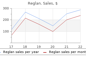
Buy 10 mg reglan. How I CURED My Chronic Illness With Celery Juice.
Moreover gastritis symptoms weight loss purchase reglan online now, the so called screw-home bursa mechanism gastritis diet �??������� discount 10 mg reglan visa, which is a combination of external rotation of the tibia occurring with knee extension gastritis x helicobacter pylori order genuine reglan line, further approximates the osseous Fibula structures and tightens the ligamentous structures to stabilize the joint (Figure 6-172) gastritis y colitis nerviosa sintomas order cheap reglan line. The marked incongruent positions of the Figure 6-170 Sagittal section through the knee gastritis symptoms spanish generic 10mg reglan free shipping, demonstrating tibiofemoral joint are reduced by the fibrocartilaginous menisci gastritis yogurt discount reglan online visa. These also help to distribute the forces of compressive loading over a greater area and reduce compressive stresses to the joint sur faces of the knee. The deep and the patellofemoral joint plays an active role in flexion and superficial infrapatellar bursae lie just under and over the patellar extension of the knee joint. Bursal sacs also lie between the semimembra ing caudal approximately 7 cm when going from full flexion to full nosus tendon and the medial head of the gastrocnemius muscle, extension. The articular surface of the patella never makes com and two bursae separate the mediolateral heads of the gastrocne plete contact with the femoral condyles; the joint space decreases mius muscle from the joint capsule. It must react to rotational forces, as well as absorb shock, In addition, it allows a wider distribution of compressive force on and then immediately prepare for propulsion. Limited rotation occurs, especially when the knee joint in both the vertical and sagittal planes. Muscle imbalance and rotational disrelationships of the tibia and Spin femur produce changes in the Q angle. Figure 6-171 Flexion and extension movements are combinations the superior tibiofibular joint is mechanically linked to the of roll, slide, and spin. The superior tibiofibular joint allows superior and inferior movement, as well as internal and external rotation of the fibula. The addition of ankle eversion causes some posterior displacement of the fibular head. Ankle plan tar flexion draws the fibula inferiorly and creates external rotation. A lateral collateral ligament injury may occur with a varus (adduction) blow to the knee, and when distance from the inferior pole of the patella to the tibial tubercle internal rotation and hyperextension occur with this force, the (Figure 6-174). To determine if the internal rotation of the femur on a fixed tibia while the knee is patella is aligned properly in the femoral groove, the Q angle can abducted and flexed. After locating the center of the patella, extend a when a traumatic force is delivered to the front of the flexed tibia, line up the center of the patellar tendon through the center point. Figure 6-175 Q angle, the angle formed from the intersection of lines from the center of the patella to the anterior superior iliac spine and After traumatic injuries to the knee causing ligamentous injury, along the quadriceps tendon. Orthopedic stress tests are performed to identify the presence of joint instability. The lumbar spine, hip, and mon and may be more frequent than ligamentous or meniscus foot are sources of referred pain to the knee (Figure 6-176). A patient complaining of vague aching pain about the To begin the evaluation of the knee, observe the knee for evi knee that is aggravated by going up or down stairs likely has patel dence of swelling, asymmetry of contours, and postural changes lofemoral joint dysfunction. Movements of the knee during gait primarily from injuries to the knee and quadriceps mechanism should be smooth and rhythmic, with the knee bent during the or secondarily in response to problems affecting the ankle or hip. Chondromalacia patella, an erosion and fragmentation of the sub Identify osseous symmetry and pain production through static patellar cartilage, may be secondary to trauma, recurrent sublux palpation of the tibial plateau, tibial tubercle, adductor tubercle, ation, pronated feet, postural instability, short leg syndrome, or femoral condyles, femoral epicondyles, fibular head, patella, and excessive femoral torsion with resultant irregular Q angle. Evaluate accessory joint motions for the knee articulations to determine the presence of joint dysfunction (Table 6-18). Assess long-axis distraction with the patient supine and the affected leg slightly abducted. Stand and face the patient, straddling the affected Figure 6-176 the ankle and hip can refer pain to the knee. Use both hands to palpate the knee joint at its medial and lateral aspects and use your legs to create a long-axis distraction while palpating with your hands for a springy end feel (Figure 6-178). Evaluate A-P and P-A glide with the patient supine and the involved knee flexed to 90 degrees with the foot flat on the table. Stress the proximal tibia in an A-P and P-A direction, looking for a springing end feel (Figure 6-179). Chapter 6 Extraspinal Techniques | 357 Figure 6-179 Assessment of anterior-to 6-179 posterior and posterior-to-anterior glide in flexion of the left tibiofemoral joint. To evaluate internal and external rotation, use the same posi tions as for A-P and P-A glide. Stress the proximal tibia internally Figure 6-181 Lateral-to-medial glide of the left and externally to feel for a springing end feel (Figure 6-180). Evaluate M-L and L-M glide with the patient supine and the involved leg abducted beyond the edge of the table. The two hands can then stress the fibula in P-A and A-P directions, looking for a springing create an M-L or L-M stress action (Figures 6-181 and 6-182). Evaluate the patellofemoral articulation for M-L glide (Figure Perform I-S and S-I glide of the tibiofibular articulation with 6-183), L-M glide (Figure 6-184), S-I glide (Figure 6-185), and I-S the patient supine, the affected leg straight, and the knee in the glide (Figure 6-186), with the patient lying supine and the involved leg relaxed extension. Use a digital contact of the cephalic hand to pal straight in passive knee extension. Figure 6-180 Assessment of external and internal rotation in flexion of the left tibiofemoral joint. Figure 6-182 Assessment of medial-to-lateral 6-182 glide of the left tibiofemoral joint. Figure 6-185 Assessment of superior-to-inferior 6-185 glide of the left patellofemoral joint. Figure 6-183 Assessment of medial-to-lateral 6-183 glide of the left patellofemoral joint. The three joints associated with the knee should be evaluated for characteristics of dysfunc tion when there are knee symptoms present. Femorotibial Joint Supine: Bimanual Grasp/Proximal Tibia with Knee Extension; Long Figure 6-186 Assessment of inferior-to-superior 6-186 Axis Distraction (Figure 6-191) glide of the left patellofemoral joint. Chapter 6 Extraspinal Techniques | 359 Figure 6-190 Assessment of inferior-to-superior 6-190 glide of the left tibiofibular joint. P: Straighten your knees while simultaneously using your hands to pull the proximal tibia into a long-axis distraction movement. Figure 6-189 Assessment of superior-to-inferior glide 6-189 of the left tibiofibular joint. Chapter 6 Extraspinal Techniques | 361 Figure 6-194 Adjustment for medial-to-lateral 6-194 glide of the left tibiofemoral joint. P: Extend your knees and hop caudal to create some long-axis distraction while simultaneously twisting the proximal tibia, either internally or externally, with your hands. Figure 6-195 Adjustment for lateral-to-medial 6-195 Hypothenar/Proximal Medial Tibia with Leg Stabilization; glide of the right tibiofemoral joint. P: Create articular tension by using your body on the distal tibia Reinforced Mid-Hypothenar (Knife-Edge)/Proximal Tibia as a lever, pivoting against the contact hand. When all joint Pull; Posterior-to-Anterior Glide in Flexion (Figure 6-196) movement is removed, deliver an M-L impulse thrust. Patellofemoral Joint Figure 6-196 Adjustment for posterior-to 6-196 Prone: anterior glide of the left tibiofemoral joint. Chapter 6 Extraspinal Techniques | 363 A B Figure 6-198 Adjustment for superior and medial (A) or inferior and lateral (B) glide of the right patellofemoral joint. P: While maintaining the ankle in eversion, deliver a thrust to the proximal fibula in an I-S direction. This movement can be achieved in a similar fashion by placing a thenar contact under the inferior aspect of the lateral malleolus, reinforc ing the contact with the other hand, and delivering an I-S thrust. Figure 6-201 Adjustment for posterior-to 6-201 Reinforced Mid-Hypothenar (Knife-Edge)/Proximal Superior anterior glide of the left tibiofibular joint in the prone position. The ankle and foot can be discussed together because they are Side Posture: intimate components of a very intricately functioning unit. Reinforced Mid-Hypothenar (Knife-Edge)/Proximal Fibula Together they make up a significant component in a kinetic Push; Inferior-to-Superior Glide in Eversion (Figure 6-202) chain responsible for propulsion and balance. The first, or medial, cuneiform articulates with the first metatarsal; the second, or intermediate, cuneiform articulates with the second metatarsal; and the third, or lateral, cuneiform articulates with the third metatarsal. Two phalanges complete the structure of the great toe, and three phalanges complete the bony structures of each of the other four toes. The function of the tibia is to transmit most of the body weight to and from the foot, and although the fibula plays a very important role in ankle stability, it is not directly involved in the transmission of weight-bearing forces. Ligamentous Structures Although many ligaments and joint capsules are associated with the foot and the ankle, some are more important to localize, palpate, Figure 6-203 Adjustment for superior-to-inferior 6-203 and functionally understand (Figures 6-205, 6-206 to 6-207). The deltoid ligament, composed of four parts, provides medial stability to the ankle by attaching from the medial malleolus to viewed as initial supports for the musculoskeletal frame, because the talus anteriorly and posteriorly, as well as to the navicular and they form the base on which all other osseous and muscular mech calcaneus. To the contrary, these joints may also be viewed as ular ligaments: the anterior and posterior tibiofibular ligaments, a terminal segment, in that they must translate and carry out the messages from the central nervous system through the hip and knee. This joint complex must attenuate weight-bearing forces, 15 Interosseous ligament support and propel the body, and maintain equilibrium. The Anterior tibiofibular ligament feet and ankles must therefore provide the two paradoxical qual Anterior talofibular ligament ities of stability and pliability. They achieve these requirements Posterior tibiofibular ligament Dorsal talonavicular ligament through an interaction of interrelated joints, connective tissues, Bifurcated ligament and muscles. Certainly, this part of the lower extremity is subject Posterior talofibular Dorsal cuneonavicular to a multiplicity of traumatic and postural disorders, leading to ligament ligament numerous joint dysfunction syndromes. The calcaneus, the larg ligament Dorsal tarsometatarsal ligament est tarsal bone, articulates with the talus, forming the subtalar joint. Interosseous talocalcaneal Dorsal calcaneocuboid the navicular articulates with the talus proximally and cuneiforms ligament ligament distally. The cuboid articulates with the calcaneus proximally and Figure 6-205 Ligaments on the lateral aspect of the right ankle. As such, it serves primarily Posterior as an inverter and adductor, or supinator, of the foot. The flexor tibiofibular hallucis longus and flexor digitorum longus muscles also have ligament tendons that pass under the medial malleolus, with each inserting on the distal phalanx of each toe, thereby creating flexion of the Transverse toes. Anteriorly are the extensor digitorum longus, tibialis ante Deltoid tibiofibular rior, and extensor hallucis longus muscles. The extensor digitorum ligament ligament longus attaches to the dorsal surfaces of each of the distal phalan ges, primarily extending the four toes, but also serving to dorsi Interosseous flex, evert, and abduct the foot. The extensor hallucis longus is the talocalcaneal primary extensor of the big toe. The tendon of the tibialis anterior ligament passes over the ankle joint and across the medial side of the dor Calcaneofibular ligament sum of the foot, and inserts into the medial and plantar surface of the medial cuneiform bone and the base of the first metatarsal. It functions as the primary dorsiflexor of the ankle, but because of its insertion, it also inverts and adducts the foot. The tendons of the peroneus Figure 6-207 Ligaments on the posterior aspect of the right ankle. The peroneus tertius is continuous, with the ori ment attaches from the sustentaculum tali to the navicular (Figure gin of the extensor digitorum longus muscle; its tendon diverges 6-208). The function of this ligament is to keep the medial aspect laterally to insert into the dorsal surface of the base of the fifth of the forefoot and hindfoot in apposition and, in so doing, help metatarsal bone. It works with the extensor digitorum longus to to maintain the arched configuration of the foot. The ankle musculature can be divided into positional groups Musculature and divided according to the actions they perform (Table 6-19). Similar to the way the muscles of the wrist are located in the arm, the gastrocnemius, soleus, and plantaris muscles lie posteriorly the muscles of the ankle are located in the calf. The large calf muscle group (gastrocnemius and soleus) attaches from extensor hallucis longus, extensor digitorum longus, peroneus the femoral condyles, proximal fibula, and tibia to the calcaneus, tertius, and tibialis anterior muscles lie anteriorly and serve pri providing plantar flexion of the ankle. The peroneus alis posterior passes under the medial malleolus to attach to the longus and brevis muscles are situated laterally and pronate and plantar surfaces of the navicular; cuneiforms; the cuboid; and the evert the foot. The ankle joint is a uniplanar hinge joint, with talus motion occurring primar Ankle dorsiflexion 20 Roll and glide ily in the sagittal plane about a transverse axis. The lateral side of the Ankle plantar 50 Roll and glide transverse axis is skewed posteriorly from the frontal plane (Figure flexion 6-209). The primary movement at the ankle mortise is dorsiflexion Subtalar inversion 5 Roll and glide (20 to 30 degrees) and plantar flexion (30 to 50 degrees) (Table Subtalar eversion 5 Roll and glide 6-20).
This would point to a gram Antibodies to the microbial products are negative bacteremia gastritis body aches generic reglan 10 mg visa. Normal C4 levels usually found in normal individuals gastritis diet 23 generic reglan 10 mg without a prescription, but with low C3 and factor B levels suggest the if they are not gastritis healing diet reglan 10mg for sale, one should suspect abnor alternative pathway alone gastritis diet ����������� generic 10 mg reglan visa. Elevation of all malities of antibody production like those three components usually suggests acute seen in immunodeciency states gastritis symptoms tagalog order on line reglan. In most cases gastritis diet ������ buy 10 mg reglan mastercard, the With the renewed interest in the role of best approach is to receive freshly biop lymphocytes in disease states over the past sied nonfrozen material that is then snap thirty years, a systematic study of the mark frozen and sections cut and stained to test ers present on B and T lymphocytes has for the presence of appropriate antigen or been undertaken. The different cell populations are rapidly due to the speed with which cells aspirated into the machine, which forces are counted with different labels, and the the cells to ow through the chamber singly results are accurate because of the large past a laser beam and light sensors. Light number of cells that are counted (in the emitted by the excited uorescent dye on thousands). It should be emphasized that the cell surface is detected by the sensors values for lymphocyte subsets will change and analyzed by computer software. Using with increasing age from children to adults, this system, one can divide cells into dif especially during the rst year of life. Finally, the num this test assumes greater importance in ber of B cells expressing a novel antigen clinical immunology both at a research is identied, in which the percentage level and in the clinical laboratories. Isolation and purication of a given subset of cells can be achieved by a uorescein-activated cell sorter. Each point on this plot represents uorescence recorded from an individual lymphocyte as it passes through the counter. The preferred to the bottom, while the mononuclear method is the isolation of the lymphocytes cell population stays in the middle of the from blood using a density gradient assay gradient. Once and slowly layered over the density gradi the mononuclear cells are removed from ent solution, which has been prepared so the gradient and washed several times, a that the cell populations will separate into relatively pure population of mononuclear different layers. The following is a short sum mary of these techniques and their impor Diluted blood tance in clinical immunology. These cells are counted lecular interaction between biological mac and adjusted to a given number of cells for romolecules. This technique may be used not only on fresh samples but also in tissues that have When these cells are stimulated by a formalin-fixed and paraffin-embedded specic antigen, a few of these resting cells tissues. In this technique of in situ hybrid undergo proliferation (activated cells) and ization, the probes can be applied directly the transformation of activated cells can be to tissue sections on microscope slides. After extensive research and handling specimens to be tested as well as workshop meetings, it became apparent in the test itself is obligatory. While most of the original testing of Dausset and others in the 1950s and for the antigens on cells and for use in 30 Immunological Techniques transplantation relied on serological tech the method is relatively straightfor niques using puried antibodies to detect ward. In addition of the human genome have been placed to the transplantation, these techniques (12,000 probe sets/chips). The chips are are used for familial genetic studies as then washed, stained with streptavidin well as for specic disease states (see phycoerythrin, and scanned with a probe other chapters in this book). Note ers will be identied in particular disease the decreased expression of red intensity in states. Most disease states appear to be polygenic, and factors such as the environment and past exposure to a given microbe also play a part in establishing the full disease pattern. Of par genomic analysis of lesional versus ticular interest, one can compare the lev nonlesional skin in psoriatic skin. The els of gene expressions across the human heat map describes 1,119 genes that have signicantly higher expression (red genome in one subset of patients in a given area) by 1. Appli bodies in metastatic prostate cancer is cations of ow cytometry to clini already well established. Flow cytometric evaluation of B diseases associated with human genetics cell lymphoid neoplasms. The other is in the treatment of the immune system in general responds autoimmune and malignant diseases. However, there are certain diseases by suppressing cytokine and chemokine that arise from either a defective or over encoding genes, which inhibits the acti responsive immune system on the part of vation and recruitment of inammatory the host. These include reader to the various approaches that have an increased susceptibility to infection, been used to either suppress or stimulate osteoporosis, and growth disturbances in the immune response. The development of the thiopurines Immunosuppressive Drugs in the 1950s ushered in a new group Several groups of drugs suppress the of immunosuppressive agents, the most immune system (see Table 3. It is the oldest of these drugs are the corticoste inactive until it is metabolized in the liver roids, which have long been known to alter and takes three to four weeks to be effec immune responses. Another major effect Another group is the alkylating agents, of in humans is on resting macrophages which cyclophosphamide is one of the best (activated macrophages are not sensitive). This drug also requires activa In humans, steroids are used for two tion by the liver. Taco by disrupting folic acid metabolism, has limus is a newer-generation drug with a similar immunomodulatory effects. Cyclosporin, a naturally occurring fungal metabolite, also inhibits T-cell acti vation and cell-mediated immunity. However, long-term tration of anti-D antibodies to the mother use has demonstrated severe toxicity such immediately after delivery. This inhibits 34 Immune Regulation the formation of anti-D antibodies in the infusions must be repeated frequently to mother, thereby avoiding the development sustain results. This thera Monoclonal antibodies can also be used peutic measure has virtually eliminated as antitumor agents (see review by Reichert). Antibodies that cancer, especially metastatic bone lesions, target the immune system can target cell and it is hoped many more monoclonal surface molecules on T or B cells or can antibodies will be forthcoming against var target soluble mediators of inammation ious other tissues. Among the most effec Other methods of immunosuppression tive uses of monoclonal antibodies has are plasmapheresis or plasma exchange. There term suppression of helper T cells and has are three principal ways to potentiate the been used in some severe autoimmune immune response in humans: through diseases like lupus or rheumatoid arthritis. Interferons bind to cell response to control chronic infections is an surface receptors and activate secondary important goal and is under active investi intracellular changes which inhibit viral gation. They can be divided into three some types of cancer can be controlled by groups: alpha , beta , and gamma 36 Immune Regulation interferons. All three interferons have pulmonary edema, and neuropsychiatric been genetically engineered, and recom symptoms. More severe effects are Examples include infusion of hepatitis B reversible: bone marrow depression, liver immune globulin and the adoptive trans dysfunction, and cardiotoxicity. Foremost is the presence rophages and is most often used in con of a clean water supply, development of ditions in which defective macrophage sanitary facilities, good nutrition, and good function occurs. More recently, immuni ders are lepromatous leprosy, leishmani zation against a particular agent has been asis, and chronic granulomatous disease. Although no vaccine ability, resulting in marked hypotension, is ideal and each has its problems, the Immune Regulation 37 problems of live vaccines are generally humans is aluminum compounds, which related to their safety, while the problems are generally safe for human use. Others of killed vaccines are related mainly to include muramyl dipeptide, biodegradable their effectiveness. However, many others are being nize in a manner similar to natural infection developed or will probably be given U. This is the Gardisal vaccine manufac now an acellular vaccine) or one of the tured by Merck to protect against human products or fractions of the organism. It is estimated the pneumococcal, meningococcal, and that ten of the thirty different serotypes Haemophilus inuenza vaccines. In general, of the virus can induce cervical cancer, so the killed vaccines are not as effective as the vaccine has been directed at eliminat the live viruses because they do not give ing those serotypes. Thus, if 10,000 women long-lasting immunity as a live infection are infected with one of the high-risk viral does. For example, although the tetanus serotypes, approximately 3,900 of them toxoid vaccine is effective, it requires a will die of cervical cancer. The biology of interleukin-2 and interleukin-15: implications for cancer therapy and vaccine design. Thus, interleukin-2 and interleukin-15: implications for cancer therapy and vaccine design. These mice develop autoimmune diseases Finally, it was shown to be effective in a such as hemolytic anemia and inamma subset of patients with adult T-cell leukemia tory bowel disease. These new devel stimulus remain to be determined and may opments are mainly based on the lessons inuence efcacy. The rst trial, published in 1998 ond, immunological monitoring of many by Nestle and colleagues, aroused great clinical trials has failed to identify a sur interest given an overall response rate of rogate marker for clinical outcomes. These ndings suggest modiers must be an essential compo the interesting possibility that the imma nent of any cancer vaccine. This approach has worked controls that act on T cells to stimulate or reasonably well with chemotherapies, inhibit them has led to the use of reagents to which, although not cancer specic, can enhance antitumor T-cell activity. For exam confer clinical benet with acceptable mor ple, blocking antibodies to the inhibitory bidities. Molecular mechanisms and a better target for breaking tolerance than cellular effects of glucosteroids. Interleukin 15: dritic cell as adjuvants for the induction biology and relevance to human dis of melanoma-specic T-cell responses in ease. Apop Interleukin-2-receptor blockade with totic cells deliver processed antigen to daclizumab to prevent acute rejection dendritic cells for cross-presentation. In general, the pool of phagocytic cells that are both cir host manages to either eliminate or ward culatory and in the bone marrow. Invad off these invading organisms, and a ing organisms trigger an inammatory symbiosis is achieved between microbes cascade, which stimulates these cells to and the host. There adhere to vascular epithelium and actively are two major pathways to achieve this migrate toward the infection. Microbes Nonspecic or natural resistance refers to that penetrate an epithelial surface will barriers, secretions, and normal ora that encounter local tissue macrophages called make up our external defenses. Once engaged with the organ exposes the host to marked susceptibility ism, these macrophages release a number to infection. The mucosal lining of mouth of macrophage-derived cytokines, which and respiratory tract is another excellent nonspecifically amplify the immuno defense mechanism. Yet, a defect in the logical and inammatory reactions to the mucosal lining of the respiratory tract, invading microbe. In gen have cell surface structures called M pro eral, however, it is the mobilization of the teins of which there are now more than phagocytic cells such as monocytes/mac 120 antigenically distinct molecules that 45 Eyes Respiratory tract Digestive system Urogenital tract Skin Figure 4. Although the structures of many dif pneumococcal polysaccharide capsule ferent pathogenic microbial compounds of which there are thirty to forty distinct have been extensively studied, the molec polysaccharides. Another approach (taken ular basis of their recognition by the cells by both group A streptococci and staphy of the innate immune system remained lococci) is the release of potent extracel elusive. Charles Janeway first devel lular toxins, which kill phagocytes with oped the concept of microbial structures the formation of pus. It has been known for decades vae) in species as diverse as Drosophila y that microbial products such as lipopoly and humans and the recognition of their 46 Immunological Aspects of Infection role in distinguishing molecular patterns is expected that many more receptors will that are common to microorganisms led be discovered in the future. The microbes display certain molecular pat importance of each arm of the specic terns that are necessary for microbial response varies from infection to infection. Many of these Experimental animal models and naturally molecular patterns such as lipopolysaccha occurring immunodeciency states clearly ride in the outer membranes of gram-nega demonstrate that certain components of tive bacteria seem to be particularly potent the immune response are crucial for con activators of mammalian cells. Yet, replacement therapy with immu lead to a delay or blunting of the immune noglobulin greatly reduces the number of response, resulting in unchecked invasion infections. It a new and rapid growth in interest in the now appears that it is not polysaccharides past eight to ten years. Since efcient phagocytosis by dent and require helper T cells for initia neutrophils requires interaction with its C3 tion of the immune response. This against these infections can be seen in is partially true if a blood sample drawn Immunological Aspects of Infection 49 Variable Conserved N A1 A2 A3 A4 A5 B1 B2 B3 B4 B5 C1 C2 C3 Pro/Gly C 600 nm Cell Wall Figure 4. The streptococcal M protein is a coiled-coil molecule that extends about 600 nm from the bacterial cell surface. The C-terminal region is embedded within the cell wall and the C-terminus is located in the cytoplasmic membrane in the nascent molecule. Pro/Gly designates the region of the M protein that is rich in proline and glycine.