
John R. Saucier, MD, FACEP
- Attending Physician, Maine Medical Center, Portland, ME
- Assistant
- Professor of Emergency Medicine, Department of Emergency Medicine
- Clinical Assistant Professor in Surgery, University of Vermont, College
- of Medicine, Burlington, VT, USA
The electronic health record data will be collected continuously over the 10 years of the study meaning participants recruited now will have 10 years of data associated with them blood pressure low symptoms discount lanoxin 0.25 mg fast delivery. There are future goals of including all parts of the electronic health record and data from wireless sensor technologies (including data from mobile/wearable devices) blood pressure diastolic low lanoxin 0.25mg on line, and also geospatial/environmental data arteria zarobki cheapest generic lanoxin uk. We conclude the All of Us Research Program discussion with some highlights and cautions regarding participation blood pressure log chart pdf buy cheap lanoxin 0.25mg on line, privacy heart attack 8 trailer buy lanoxin visa, and access blood pressure bottoming out order generic lanoxin on line. All of Us Research Program Participation, Privacy, and Access A major purpose of the All of Us Research Program will be to make the data available to participants, researchers, and the public. To build confidence around privacy, All of Us Research Program is laying the foundation for ensuring confidentiality, integrity and availability of the data. This is through the activities of two interagency working groups tasked with developing privacy and trust principles and data security policy principles and framework. Participants may not want to share all of the required information and so a mechanism must be put into place to enable participants to choose which pieces of information can be released and to whom. On the one hand, novel methods to help participants understand informed consent are being developed based on research that has 37 been piloted by Sage Bionetworks [118]. These methods have now become part of the All of Us Research Program protocol [119]. The informed consent process will allow all participants to opt-in to all or only subsets of their data being included in the study and/or shared with research studies. Participants will also be able to change their preferences and opt-out at any time. On the other hand, parallel consent authorization processes need to be implemented properly. For example, one may want to access a web service without being a locally authorized user of that service. In some cases, the service will offer to authenticate the user through their credentials used for another site typically a large provider. The remote site then authenticates the user and may also query the use for which authorization is requested. If the user accedes a token is sent to the requesting site indicating the user is valid and has given permission to the site to authenticate them. From then on when the user wants access to the web service the authentication can be mediated through the remote provider. This approach makes it possible to create web services that can consolidate disparate pieces of data in one place. A common example is a site to let someone see their comprehensive financial position by interrogating all the financial service sites in which a user participates and providing a composite analysis of the data. In the past, this required having the user provide authentication data to the consolidation site so that it could perform remote queries. Because no authentication information is provided the consolidation site can only query the limited piece of information. The exchanged tokens encode the level of permissions, so if a user does not want to give access to some account data, it is at their discretion not to do so. If the user decides not to share the information the permissions can always be revoked, and the tokens then become invalid. For example, ones privacy policies for research projects may differ from those associated with an insurance company. Or one may allow data associated with blood tests to be released but may refuse to share data associated with mental health. Some of these have suffered security breaches because the protocol was not implemented properly. It may be necessary to have a secondary verification such as confirmation on ones cell phone to verify that a request is valid. It is not easy to decompose the pages into the atomic data elements associated with a privacy bundle. The second S4S technology is the use of blockchains as a way of validating the provenance of data. Because the All of Us Research Program will be highly distributed, the problem arises that various pieces of data may become out of sync with one another. One way to deal with this is to have a central point of access for the data but this may lead to significant inconvenience on the part of researchers who are distributed around the country. It would also create a single point of failure should the central repository suffer an interruption of service either due to a security incident or perhaps hardware failure. The idea of the blockchain is to create a distributed ledger where various accesses or changes in the ledger are indicated through the use of a set of interlinking hash codes (known as a Merkle tree [78]). Using this type of data structure, it is possible to verify who made a change and what change was made. It is virtually impossible to corrupt the database because a change in the data causes changes in the hash codes. While it is theoretically possible to forge data with the same hash codes, to date there are no algorithms known that can do this in a reasonable period with current computing resources. In addition, even if one could forge the data, because the database is distributed and available to everyone it is virtually impossible to insert forgeries on all the copies. There is a special concurrence algorithm that allows all users to eventually learn which blockchain is the most recent making it unnecessary for all users of the ledger to perform specific synchronization of their copies. The use of blockchains would be particularly appropriate for health records in general. It solves the problem of trying to locate the most current records when a patient presents themselves at a 39 medical facility. By querying the blockchain all interactions would be recorded and the most recent data could be acquired. Agreeing to the use of this technology means that a patient is tracked throughout the health care system. One type of environmental exposure that is closely monitored by epidemiologists and clinicians is pathogen exposure. It is interesting to think that someone might live right next to an industrial smokestack, yet this may never make it into their medical record because clinicians dont typically ask questions related to environmental toxicology, and these have not been incorporated into most standard health questionnaires. For many diseases, environmental exposures play a bigger role in health outcomes than genetics. Yet, the amount of attention paid to environmental factors is a fraction of the attention that has been devoted to genetics. There are well substantiated environmental risk factors for significant diseases such as many cancers [124] and autism [125,126], yet to our knowledge these known environmental components are not being tracked as part of most health data projects. Data in all of these areas should continue to be developed, but we find that absence of the environmental exposure datastream to be particularly troubling. For instance, one person might have many pieces of furniture containing flame retardants [127], which have been linked to cancer, while another does not. At some level, this information can also be captured by measuring toxins in individuals bloodstreams. One can imagine that members of some households may have higher levels of certain toxins than members of another household. Environmental exposure data can fairly easily be captured in health data projects. For instance: Blood testing could include assays for common environmental toxins. Custom and commercial testing kits are available (for example [128] Diet-related toxins should be considered. Patient questionnaires could be designed to include 40 questions about diet-related toxins, for instance the percentage of produce purchased that is organic, number of times per week that seafood [129] is consumed, etc. There is some evidence that, in certain places in the country, water contaminants including lead fall outside of allowable bounds [131]. Collection of parallel city-scale environmental data should be incorporated at health study sites (for instance, the cities where the All of Us Research Programs data collection centers are located). This can be done by city-wide sensors, as described in the next section, or by having participants wear or carry devices that can take these measurements. These are important data sources to be used in building an understanding of the social determinants of health and the impact of the environment on health, but a finer level of geographic resolution in the capture of environmental exposure data likely will be needed to unravel the relationship between health status, genetics, environment and behaviors. There are numerous academic projects underway to collect environmental data within urban settings. Indeed, there is a flourishing of academic and for-profit efforts to measure cities and exploit the data so derived [135]. For instance, the Array of Things project is a network of urban sensors placed on telephone poles around Chicago [136] and the Sounds of New York City project [137] aims to measure and characterize the urban noise field with high spatial and temporal granularity through stationary in-situ sensors. Urban metagenomic studies are underway to characterize the space-time variation of the urban microbiome and light pollution in a given housing unit can be measured passively and synoptically by observations of building facades from urban vantage points. The fusion of such data with individual mobility tracks can provide individual exposures. Such environmental data should be collected as part of all big-data health and health care projects. Collection of these data streams is particularly practical in projects that are based out of specific medical centers, limiting the environmental data needed to a few particular cities. Studies should also be designed to place sensors within the homes of individuals, with care for privacy concerns, so that the home-to-home variability in environmental exposure can be evaluated. Recommendation: Support ambitious and creative collection of environmental exposure data. The perils of this assumption, and the associated need to address it with systematic approaches to both data management and transparency in algorithm development is discussed in this section. In the context of deep learning, understanding the basis of a models output is particularly important as deep learning models are unusually susceptible to adversarial examples and can output confidence scores over 99. In addition, the issue of interoperability of electronic health record systems between care settings remains elusive [142]. The study addressed a serious issue, which is that standard assessments do poorly in predicting the patients 43 who eventually do have a cardiovascular event, and generate huge numbers of false positives that stymie effective follow on testing. The potential to improve risk assessment using machine learning was assessed using electronic health records for the time period between 2005 and 2015. From the 12 million patient records, about 375,000 were suitable for use based on the requirement of complete records on 8 standard diagnostic indicators. Three quarters of the records were used as the training case for machine learning, and other quarter were used as the test case. In addition, for the machine learning assessments, 22 more diagnostics available in the health records were added to the input data streams. The statistical results for the standard risk tool and the two best performing machine learning algorithms are summarized in Table 4. The machine learning algorithms improve the sensitivity (true positives) by almost 5%, but increase the specificity (decrease the false positives) by less than 0. Given the poor baseline, the improvement in sensitivity still leaves much to be desired in correctly identifying patients at risk. The very small improvement in specificity yields an insignificant impact on the serious issue of false positives. Another possibility is that there may be errors in the data used for the training set. Examples of the sensitivity and specificity of the health records relative to the research study measurements include: Hypertension sensitivity: 71. The changes in the ranked risk factors for the two best performing machine learning algorithms appear almost idiosyncratic, consistent with the well-known black-box nature of machine learning. This is independent of the hope that there will be the kind of continued improvements that have occurred in image recognition or various aspects of natural language processing. Another observation is that not all errors are equally important (or unimportant). As the system is being developed one typically uses error curves or recall/precision statistics, and without special treatment these evaluate all errors as the same. It is easy to imagine similarly unexpected, but possibly life-threatening errors in health applications. Assessment of algorithms must include questioning whether the observed error rates are like the expected rates, and identifying what types of errors the algorithm makes and why. Sometimes the changes are relatively slow, as with the multi-decadal change in the kinds of pneumonia seen [148]. Sometimes new diseases pop up and require changes to previously sound diagnostic protocols. Thus, even if an application of deep learning were ideally suited to the real world when it is first released, over time the real world may drift and make a static application less and less effective. Assessment of algorithms should include understanding how they will respond, or what indicators may be observed, if the input data characteristics begin to diverge from the original training sets. There has been recognition that guidance on reproducibility for computational methods is needed. Stodden [149] points out there are actually three important parts to consider: empirical reproducibility, computational reproducibility, and statistical reproducibility. Empirical and statistical are being readily addressed by the research community, however computational is lacking. They note: Over the past two decades, computational methods have radically changed the ability of researchers from all areas of scholarship to process and analyze data and to simulate complex systems. But with these advances come challenges that are contributing to broader concerns over irreproducibility in the scholarly literature, among them the lack of transparency in disclosure of computational methods. They recognize that meeting these principles will be challenging and exceptions will be necessary with human subject research and proprietary codes.
Results were reported separately for kidney cancer (n = 186 cases) and renal pelvis cancer (n = 23 cases) heart attack 99 blockage lanoxin 0.25 mg fast delivery, but no excess cancer risk for the kidney or renal pelvis was found when compared with the general Korean population (Yi blood pressure medication used for anxiety order lanoxin toronto, 2013) or when internal comparisons of highversus low-exposure-opportunity scores were made (Yi and Ohrr arrhythmia with pacemaker cheap lanoxin 0.25mg line, 2014) arterial blood gas buy lanoxin 0.25 mg on line. When kidney pulse pressure mayo clinic lanoxin 0.25 mg discount, renal pelvis heart attack xbox cheap lanoxin 0.25mg with mastercard, and ureter cancer deaths were combined for the internal cohort comparison of high versus low exposure, no excess cancer mortality was found (Yi et al. Information on smoking or other lifestyle habits was not available for this cohort during the follow-up through 2003, and thus the modest associations could be due to confounding by smoking or obesity. No studies of renal cancers in Vietnam veterans have been identifed since Update 2014. Studies of Vietnam veterans have not found statistically signifcant associations between deployment and presumed exposure to the herbicides and incidence or mortality of renal cancers. Similarly, no increases of risk or mortality from renal cancers have been reported among the several occupational cohorts, where exposure was often better characterized. Glioblastoma multiforme is the most common brain tumor and has the worst prognosis (M uth et al. Several types of cancer are usually grouped together; although this may bias results in unpredictable ways, the most likely consequence is a dilution of risk estimates toward the null. The causes of most cancers of the brain and other portions of the nervous system are unknown. The committees responsible for Update 1996, Update 1998, Update 2000, Update 2002, and Update 2004 did not change that conclusion. That committee considered one study that suggested a relationship between phenoxy acid herbicides and adult gliomas (W. The committees for Update 2008, Update 2010, and Update 2012, reviewed several new occupational, environmental (including updates of the Seveso cohort), case-control, and Vietnam veteran studies, but maintained that brain cancer should remain in the inadequate or insuffcient category, given the largely null fndings and that several studies did not specify the chemicals of exposure. Vietnam veterans nurses study is limited by the issue of multiple comparisons, the possibility of false positives, and imprecise risk estimates. Update of the Epidem iologic Literature Because glioblastoma multiforme was specifcally noted as an outcome of importance in the committees statement of task, a targeted literature search for this outcome was undertaken. No date or language parameters were applied, and a total of 153 articles were found. Each case was frequency matched to four controls, white males diagnosed with other cancers who were randomly selected from each of six age strata and information on occupation (usual or longest held) and tobacco smoking history (never, former, current). Additional analyses were conducted for specifc brain cancers, including unspecifed astrocytomas, unspecifed glioblastomas, anaplastic astrocytomas, and other (oligodendrogliomas, unspecifed cell types, and unspecifed ependymomas). When cases and controls were compared by tobacco smoking status, no difference in risk for brain cancer was found. The study is limited by the lack of specifc exposure information; industry and occupation information was incomplete in the registry (of the initially eligible subjects, 34% of cases and 38% of controls were excluded from the fnal sample due to missing information), restricted to usual or longest held job, and obtained from the medical record at the time of diagnosis and subject to misclassifcation. Therefore, while these data are consistent with some other studies that suggest an agricultural chemical exposure risk for brain cancer, they are very nonspecifc and must be considered exploratory. Hearing from the scientifc experts ensured that the committee had information that was as complete and current as possible regarding the science of glioblastoma, whereas hearing from the families of veterans who had been diagnosed with glioblastoma provided a reminder of the burden of the disease. The committee appreciates the efforts of all presenters and members of the public who attended the open session and their willingness to provide information for the committees consideration. Although a relatively small number of all new cancer cases each year originate in the brain or nervous system for both men and women (1. Gliomas are the most common type of malignant brain tumor, accounting for approximately 26. There are multiple subtypes of glioma, with glioblastoma being the most common (56. In the United States, incidence is about 50% higher in males than in females, highest for non-Hispanics whites, and associated with higher socioeconomic status. This review highlighted the stark fact that there has been little change in the incidence rate of glioblastoma since the 1990s and little progress eliciting clear risk factors. Of interest, about 25% of glioma risk is estimated to be genetic, and current research has identifed 12 common genetic variants that explain approximately 27% of the genetic risk for glioblastoma. Accepted non-genetic risk factors include exposure to ionizing radiation and a history of respiratory allergies and atopic disease, specifcally asthma and eczema. While associations with other exposures have been studied, including herbicide exposure, no additional accepted risk factors (including immune suppression arising from various exposures) have been found. These novel mechanisms may fundamentally change how we think about the evolution of this (and other) cancers. At the same time, this understanding of the basic biology has so far not directly led to new treatment options. The planned studies included an update of the causes of mortality of deployed and Vietnam era veterans from 1979 through 2014 and an exploratory study of self-reported exposures and different types of brain cancer using information, in part, from the Agent Orange Registry. One such effort discussed was Sierra Valley Cancer Registry Services, which is a registry that collects self-reported information related to exposures and confrmed diagnosis of glioblastoma for Vietnam veterans. As of 2017, the registry contains information on 372 Vietnam veterans who have been diagnosed with glioblastoma. This correlation was confrmed in a further study of 14 human neuroblastoma samples. Synthesis Studies of Vietnam veterans have not found statistically signifcant associations between deployment and presumed exposure to the herbicides and incidence or mortality of brain or other nervous system cancers. However, the study lacked exposure estimates and was underpowered and potentially biased by missing data, and, ultimately, the committee considered it an exploratory analysis and did not give it full weight. Given the limited epidemiologic data available on glioblastoma, the committee heard invited presentations from two glioblastoma experts. The thyroid contains two main types of cells: follicular cells, which synthesize and store thyroid hormones and synthesize thyroglobulin, and C cells, which synthesize the hormone calcitonin, which regulates calcium metabolism. The four main types of thyroid cancer are papillary cancer, follicular cancer, anaplastic cancer, and medullary carcinoma (W iltshire et al. Papillary carcinoma is the most common and accounts for the majority of the increasing incidence rate (Lubitz and Sosa, 2016). Follicular carcinoma (or follicular adenocarcinoma), which is associated with inadequate dietary iodine intake, accounts for about 10% of all cases and has greater rates of recurrence and metastasis. M edullary carcinoma, a cancer of the parafollicular cells in the thyroid, is less common (4% of all cases) and tends to occur in families. As radiation exposure is recognized as a risk factor for thyroid cancer, increased incidence is being observed in people who received radiation therapy directed at the neck (a common treatment in the 1950s for enlarged thymus, adenoids, and tonsils and for skin disorders) or who were exposed to iodine-125, for example, from the Chernobyl nuclear power-plant accident. If the radiation exposure occurred in childhood, then the risk of thyroid cancer is further increased. Analysis of incidence and mortality of cancers in the Korean Veterans Health Study was reviewed in Update 2014. There were no statistically signifcant differences in the incidence of or death from thyroid cancer when compared to the general Korean population. Nor were there differences between the highand low-exposure groups (Yi and Ohrr, 2014). However, based on 11 deaths, a statistically signifcant association between exposure and thyroid cancer-specifc mortality was found both when analyzed in terms of log increments in the exposure opportunity scores and when comparing highversus low-exposure groups (Yi et al. The small number of cases and imprecise estimates did not change the conclusion that there was inadequate or insuffcient 18As calculated on the site seer. No pathology was available, and no clinical information on the patients was reported. A total of 19,592 thyroid cancer cases were identifed, 42% of which were among Vietnam-era veterans. The authors found a statistically signifcantly higher proportion of self-reported Agent Orange exposure among thyroid cancer patients (10. However, this analysis is limited by the absence of pathology reviews of identifed cases, no reporting of histological subtypes, and no adjustment or inclusion of additional information on comorbidities or other risk factors. W ith limited deaths, mortality risk estimates were imprecise and not statistically signifcant for any of the groups of workers. There was a signifcant increase in follicular-cell adenoma in female but not in male mice. Thus, although human and animal studies showed that dioxin and dioxin-like chemicals alter thyroid hormones and increase follicular-cell hyperplasia, there is little evidence of an increase in thyroid cancer. There are some reports of therapeutic treatment with arsenic trioxide and later development of thyroid cancer (Au et al. As indicated in Chapter 4, 2,4-D and 2,4,5-T are at most weakly mutagenic or carcinogenic, and no studies that addressed a possible association between exposure to those herbicides and thyroid cancer in animal models have been identifed. They are among the most common types of cancer induced by environmental and therapeutic agents. The categorization of cancers of the lymphatic and hematopoietic systems has changed over time, guided by growing information about somatic mutation, gene expression, and subclonal lineage of the cancer cells that characterize each of a broad spectrum of neoplasms arising in these tissues (Jaffe, 2009). This classifcation was updated in 2016 and reviewed by several academics and clinicians (Arber et al. Stem cells arising in the bone marrow generate two major lineages of leukocytes: myeloid and lymphoid. M yeloid cells include monocytes and three types of granulocytes (neutrophils, eosinophils, and basophils). Lymphoid cells include T and B lymphocytes and a smaller set of cells called natural killer cells. All of these mature cells circulate in the blood and are collectively referred to as white blood cells or leukocytes. Monocytes move out of the bloodstream into infamed tissues, where they differentiate into macrophages or dendritic cells. Antigen stimulation induces the T cells to differentiate into several subtypes involved in cell-mediated immunity, immune regulation, and the facilitation of B cell function. Progenitor or pre-B cells mature in the bone marrow into antigen-specifc B cells. On encountering their cognate antigens, B cells differentiate into antibody-secreting plasma cells involved in humoral immunity. The normal cells are transformed into a malignant cell population through a multistep process that involves genetic and epigenetic alterations. Leukemias occur when a myeloid stem cell residing in the bone marrow becomes transformed, resulting in a failure of differentiation and a resistance to normal feedback on cellular proliferation. As the leukemic cells (blasts) fll the bone marrow, they actively secrete cytokines that prevent normal cellular proliferation, leading to reduced circulating normal blood cells. In addition, changes in adhesion molecules allow the release of these immature cells into the peripheral blood. Leukemias are generally classifed as myeloid or lymphoid, depending on the lineage of the malignant cell population. Lymphoma is a general term for malignancies that arise from lymphocytes (B, T, or natural killer cells). Lymphomas generally present as solid tumors at lymphoid proliferative sites, such as lymph nodes and the spleen. As stem cells mature into B or T cells, they pass through several developmental stages, each with unique functions. About 85% of lymphomas are of B-cell origin, and 15% are of T-cell or natural killer-cell origin (Jaffe et al. B cells give rise to a wide array of neoplasms, which are characterized by the stage at which B-cell development was arrested, as well as by the surface protein expression and the genetic characteristics of the malignant cells. M ultiple myeloma is a lymphohematopoietic malignancy derived from antibody-secreting plasma cells, which also have a B-cell lineage, that accumulate primarily in the bone marrow but may also infltrate extramedullary sites. It represents a substantial advance in understanding the biologic paths by which these malignancies develop. Furthermore, the existing records that will serve as the basis of many current and even future studies will use earlier and evolving classifcations, so a confounding of classifcation is likely to remain, even in new literature. The nomenclature has become more uniform in recent studies, but the possibility of ambiguity remains if earlier researchers did not use a unique code in accordance with some established system. The Update 2014 committee familiarized itself with the classifcation systems that have been used for lymphoid malignancies, including hearing a presentation from the International Lymphoma Epidemiology Consortium (InterLymph) describing a proposed classifcation of these cancers into subtypes that are particularly appropriate for epidemiologic research, including methods to harmonize data, standardized defnitions of disease entities and rigorous quality control of these subtype assessments, and attempts to understand the implications of etiologic heterogeneity (M orton et al. Furthermore, treating these cells with benzo[a]pyrene suppresses B-cell differentiation. Data on human hematopoietic stem cells and from the use of knockout Ahr mouse models show that Ahr is critical in hematopoietic stem cell maturation and differentiation (Ahrenhoerster et al. They did note a non-signifcant decrease in most lymphocyte subsets, which was most prominent for B cells. No new mechanistic or biologic plausibility studies regarding lymphohematopoietic cells have been identifed by the committee since Update 2014. In addition to the occupational associations discussed below, higher rates of the disease have been observed in people who have suppressed or compromised immune systems. Additional studies available to the committees responsible for subsequent updates have not changed that conclusion. Several of the other case-control and occupational-cohort 19As calculated on the site seer. Other populations of Vietnam-era veterans likewise did not fnd an association (Anderson et al.
Obesity (or being very overweight) is known to increase a mans risk of dying from prostate cancer arrhythmia test discount lanoxin american express. Eating right blood pressure medication benicar side effects order 0.25 mg lanoxin fast delivery, exercising arrhythmia beta blocker cheap lanoxin 0.25mg amex, watching your weight and not smoking can be good for your health and help you avoid prostate cancer blood pressure medication not working lanoxin 0.25mg on-line. Some healthcare providers believe drugs like finasteride (Proscar ) and dutasteride (Avodart ) can prevent prostate cancer hypertension and pregnancy generic lanoxin 0.25 mg with visa. Studies do show that men taking these drugs were less likely to be diagnosed with prostate cancer arteria magna buy 0.25mg lanoxin fast delivery. Still, it is not clear if these drugs are affective so you should talk to your doctor about the possible side effects. To find out if prostate cancer screening is a good idea, take our Know Your Stats Risk Assessment Test. Tell your results to your healthcare provider when you talk about the benefits and risks of. For this exam, the healthcare provider puts a lubricated gloved finger into the rectum. During this test, the doctor feels for an abnormal shape or thickness to the prostate. When found early, it can be treated early which helps stop or slow the spread of cancer. Or, the test may be a "false positive," suggesting something is wrong when you are actually healthy. The test might also detect very slow growing cancer that will never cause problems if left untreated. For a prostate biopsy, tiny pieces of tissue are removed from the prostate and looked at under a microscope. The pathologist is the doctor who will look carefully at the tissue samples to look for cancer cells. Your doctor will also consider your family history of prostate cancer, ethnicity, biopsy history and other health factors. Prostate biopsy is usually done using an ultrasound probe to look at the prostate and guide the biopsy. Your healthcare provider will note the prostate glands size, shape and any abnormalities. The prostate gland is then numbed (anesthetized) with a needle passed through the probe. Then, the provider removes very small pieces of your prostate using a biops device. If cancer cells are found, the pathologist will assign a "Gleason Score" which helps to determine the severity/risk of the disease (see Stages for more information). Grading (with the Gleason Score) and staging defines the progress of cancer and whether it has spread: Grading When prostate cancer cells are found in tissue from the core biopsies, the pathologist "grades" it. The grade is a measure of how quickly the cells are likely to grow and spread (how aggressive it is). With this system, each tissue piece is given a grade between three (3) and five (5). A high grade of five (5) indicates a highly aggressive, high-risk form of prostate cancer. The Gleason system then develops a "score" by combing the two most common grades found in biopsy samples. The highest score of grades 5 + 5 = 10 means that cancer is present and extremely aggressive. The Gleason score will help your doctor understand if the cancer is as a low-, intermediateor high-risk disease. If you are diagnosed with prostate cancer, ask about your Gleason score and how it impacts your treatment decisions Staging Tumor stage is also measured. Staging describes where the cancer is within the prostate, how extensive it is, and if it has spread to other parts of the body. The imaging tests show if and where the cancer has spread, for example: to lymph nodes or bone. It may spread to the nearby seminal vesicles, the bladder, or further to the lymph nodes and the bones. Many men with prostate cancer will not die from it; they will die from other causes. Survival rates for men with prostate cancer have increased over the years, thanks to better screening and treatment options. Today, 99% of men with prostate cancer will live for at least 5 years after diagnosis. One out of three men will survive after five years, even if the cancer has spread to other parts of the body. Before you decide what to do, you should consider how immediate and long-term side effects from treatment will affect your life, and what youre willing to tolerate. With more experienced surgeons, the risk of permanent side effects (like incontinence) is lower. Eating a well-balanced diet, maintaining a healthy weight, exercising and not smoking are all important factors when fighting prostate cancer. Treatment choices for prostate cancer include: Surveillance Active Surveillance Watchful Waiting Localized Therapy Surgery Radiation Therapy. Cryotherapy Focal Therapy Systemic Therapy Hormonal Therapy Chemotherapy Immunotherapy Updated August 2018 Clinical Trials Clinical Trials Clinical trials are research studies involving real patients to test if a new treatment or procedure is safe, effective and maybe better than established options. The goal is to learn which treatments work best for certain illnesses or group of people. To search for information on current clinical trials for the treatment of bladder cancer, visit the UrologyHealth. Prostate cancer can be a manageable disease if caught early and treated appropriately. Talk to your healthcare provider about the side effects or problems you have after treatment. If you havent yet started treatment, consider the expertise of your doctor before you begin. With more experienced surgeons, the risk of permanent side effects, like incontinence, is lower. Whatever youre feeling, its important to tell your healthcare provider about it. Build a plan with your provider or a counselor to deal with your emotional health and general wellbeing. Erectile dysfunction and urinary incontinence are the side effects reported most often by men following prostate cancer treatment. An erection happens when sexual arousal causes nerves near the prostate to send signals. In addition, the amount of blood flowing to the penis can decrease after treatment. While most surgeons try to perform a nerve sparing procedure, it is not always possible. It may take up to 24 months or longer before you are able to have a full erection, but it is possible. Even with nerve-sparing surgery, erections do no return right away or to full pre-surgery function. They include pills, vacuum pumps, urethral suppositories, penile injections and penile implants. They can help you decide which individual or combination of treatments is right for you. Incontinence Issues After Prostate Cancer Treatment Incontinence can sometimes result from treatment. After prostate cancer treatment, you may experience different types of Incontinence. Because incontinence may affect your physical and emotional recovery, it is important to understand how to manage this problem. If you have stress incontinence (the most common type after surgery), you may need to wear a pad for a few weeks to months. Physical therapy focused on the pelvic floor may help you recover bladder control sooner. Treatment for incontinence depends on the type and severity of the problem: Kegel Exercises strengthen your bladder control muscles. Lifestyle Changes include modifying your diet, no longer smoking, losing weight and timed visits to the bathroom can decrease urination frequency. Medication affect the nerves and muscles around the bladder, helping to maintain better control. Surgery to inject collagen to tighten the bladder sphincter, implanting a urethral sling to tighten the bladder neck, or an artificial sphincter device used to control urination. Products There are also many pads and products available that do not treat incontinence but help maintain a higher quality of life. If youd like to learn more about how to manage advanced prostate cancer, read our advanced prostate cancer article. Updated August 2018 More Information More Information Questions to Ask Your doctor Diagnosis: What is my Gleason score, the grade and the stage of my cancer Treatment: What are my treatment choices (including surveillance, localized therapy or systemic therapy) What are the chances that my cancer will return after treatment and if it does, what options for treatment do I have then Side Effects & Recovery: What are the potential side effects of the treatment you recommend: both immediately and in the long term Will I need to take time off from work or other activities to manage treatment and treatment side effects Baker Family Foundation Mary Ann and Bill Becker Precision is the George and Mary Nell Berry Dr. Chalsty the Deeks Family Foundation Youll read a lot about precision in this issue of Discovery: Precision diagnosis, precision imaging, precision treatment of localized disease and precision oncology R. There is so much to tell you about, in prostate cancer, and in other cancers, as well. More precision diagnoses may be on the way with a liquid Virginia and Warren Schwerin biopsy for bladder cancer. And in kidney cancer, the American Urological Donald and Susan Sturm Association has changed its guidelines in large part due to the work of a Brady team Carolyn and Bill Stutt led by Mohamad Allaf and Phillip Pierorazio. Thats our goal for every patient we see here at the Brady: Luciana and Joe Vittoria to fnd the best treatment for that persons specifc disease. Best wishes, For additional news and updates from the Brady Institute, please follow us on our web site at urology. Twitter: @brady urology; the Jakurski Family Director Urologist-in-Chief bradyurology. Theyre Or, as Pienta puts it, Everything we are more likely to be cured than ever. Several years ago, Brady investigator thought that 70 to 80 percent of men had Not very, and the Brady team discovered Martin G. Coffey Professor of Urology, the investigators used markers for frustrated scientists for decades. In the Professor of Oncology, and Professor of epithelial cells (cells in the lining of past, scientists tried to engineer antibodies Pharmacology and Molecular Sciences, tissue and organs), which were not to recognize prostate-specifc molecules, which is really a scary number. There are different the antibody doesnt work well with lots of disease who light up with checkpoints, and new drugs that target and generally falls off in the liver. Pienta is a pioneer in the study that the side effects are fewer with one of macrophages in cancer, and is invesTreating Oligometastatic Cancer type than with another. Pienta co-led tigating and designing new therapies to a think tank meeting with scientists Brady physicians and scientists are at knock out these macrophages. This is called oligometastasis, better in combination with immunoBrady, says Pienta. If a spot of cancer has a diabolical way of putting the please use the contribution card included does show up in the bone, it can be bodys great immune warrior cells, T in this issue or contact Elissa Kohel treated focally, as well, with stereotactic cells, to sleep. Youve done it for so long, and thank goodlonger periods of time, says Patrick C.
The inability to take in enough calories to provide nutrients for thermogenesis and growth 7 pulse pressure readings buy lanoxin 0.25 mg cheap. Premature infants subjected to acute hypothermia respond with peripheral vasoconstriction arteria jugularis externa lanoxin 0.25mg low price, causing anaerobic metabolism and metabolic acidosis arteria facialis linguae best order for lanoxin. This can cause pulmonary vessel constriction heart attack 43 year old woman buy generic lanoxin 0.25mg line, which leads to further hypoxemia pulse pressure refers to buy lanoxin on line, anaerobic metabolism pulse pressure wave order lanoxin 0.25 mg online, and acidosis. Premature infants are therefore at great risk for hypothermia and its sequelae. The more common problem facing premature infants is caloric loss from unrecognized chronic cold stress, resulting in excess oxygen consumption and inability to gain weight. It occurs more often in home deliveries, emergency deliveries, and settings where inadequate attention is paid to the thermal environment and heat loss. These infants may have a bright 178 General Newborn Condition 179 red color because of the failure of oxyhemoglobin to dissociate at low temperature. Signs may include the following: (i) hypotension; (ii) bradycardia; (iii) slow, shallow, irregular respiration; (iv) decreased activity; (v) poor sucking reex; (vi) decreased response to stimulus; (vii) decreased reexes; and (viii) abdominal distention or vomiting. Metabolic acidosis, hypoglycemia, hyperkalemia, azotemia, and oliguria are present. Placing newborns in sunlight to control bilirubin is hazardous and may be associated with signicant hyperthermia. If environmental temperature is the cause of hyperthermia, the trunk and extremities are the same temperature and the infant appears vasodilated. Wet infants in the delivery room are especially susceptible to evaporative heat loss. This is a minor mechanism of heat loss that occurs from the infant to the surface on which he or she lies. Thermoneutral conditions exist when heat production (measured by oxygen consumption) is minimum and core temperature is within the normal range (Table 15. A cap is useful in preventing signicant heat loss through the scalp, although evidence suggests that only caps made of wool are effective. Examination in the delivery room should be done with the infant under a radiant warmer. Additional interventions during the rst 10 minutes can optimize thermoregulation. External heat sources, including skin-to-skin care and transwarmer mattresses, have demonstrated a reduction in the risk of hypothermia. These infants should be placed in a polyethylene bag immediately after birth; the wet body is placed in the bag from the neck down. Plastic wraps and plastic caps also have been effective in infants born at less than 29 weeks. Alternatively, skin mode or servo control can be set so that the incubators internal thermostat responds to changes in the infants skin temperature to ensure a normal temperature despite any environmental uctuation. Humidication of incubators has been shown to reduce evaporative heat loss and decrease insensible water loss. Risks and concerns for possible bacterial contamination have been addressed in current incubator designs, which include heating devices that elevate the water temperature to a level that destroys most organisms. Notably, the water transforms into a gaseous vapor and not a mist, thus, eliminating the airborne water droplet as a medium for infection. Servocontrolled open warmer beds may be used for very sick infants when access is important. The use of a tent made of plastic wrap or barrier creams such as Aquaphor (or sunower seed oil in developing countries) prevent both convection heat loss and insensible water loss (see Chap. Double-walled incubators not only limit radiant heat loss but also decrease convective and evaporative losses. Current technology includes the development of hybrid devices such as the Versalet Incuwarmer (Hill-Rom Air-Shields) and the Giraffe Omnibed (Ohmeda Medical). They offer the features of both a traditional radiant warmer bed and an incubator in a single device. This allows for the seamless conversion between modes, which minimizes thermal stress and allows for ready access to the infant for routine and emergency procedures. Premature infants in relatively stable condition can be dressed in clothes and caps and covered with a blanket. Heart rate and respiration should be continuously monitored because the clothing may limit observation. A servocontrolled warmer can generate excess heat, which can cause severe hyperthermia if the probe becomes detached from the infants skin. Servo control of temperature may mask the hypothermia or hyperthermia associated with infection. A record of both environmental and core temperatures, along with observation for other signs of sepsis, will help detect infections. Body weight and input and output should be closely monitored in infants cared for on radiant warmers. Heat loss prevention: a systematic review of occlusive skin wrap for premature neonates. Interventions to prevent hypothermia at birth in preterm and/or low birthweight infants. Fortunately, the rate of very preterm births appears to have stabilized after a persistent increase over the period from 1990 to 2005; associated with the rising twin and triplet rate presumed to be related to increased use of fertility therapies. Admissions during the rst year of life are most commonly for complications of respiratory infections. In a recent study of extremely premature infants, 57% of infants born between 23 to 25 weeks gestation and 49% of those born between 26 to 28 weeks required rehospitalization in the rst 18 months of life. Likewise, good hand hygiene by all those in close contact with infants, avoidance of exposure to others with respiratory infections (especially young children during the winter season), and avoidance of passive cigarette smoke exposure to prevent illness caused by respiratory viruses should be recommended to families. Medically stable, thriving infants should receive the Hepatitis B vaccine as early as 30 days of age regardless of gestational age or birth weight. If the baby is ready for discharge to home before 30 days of age, it can be given at the time of discharge to home. Although studies evaluating the long-term immune response to routine immunizations have shown antibody titers to be lower in preterm infants, most achieve titers in the therapeutic range. Many of these infants also have abnormal or delayed oral motor development and have oral aversion because of negative oral stimulation during their early life. Growth should be followed carefully on standardized growth curves using the childs age corrected for prematurity for at least the rst 2 years of life. Specialized premature infant formulas with increased protein, calcium, and phosphate (either added to human milk or used alone) should be considered in the rst 6 to 12 months of life in infants who have borderline growth. However, if their growth runs parallel to the normal curve, they are usually demonstrating a healthy growth pattern. Infants whose growth curve plateaus, or whose growth trajectory falls off, warrant further evaluation to assess caloric intake. If growth failure persists, consultation with a gastroenterologist or endocrinologist to rule out gastrointestinal pathology, such as severe gastroesophageal reux disease, or endocrinologic problems, such as growth hormone deciency, should be considered. Gastrostomy tube placement may be necessary in a small subset of patients with severe feeding problems. Long-term feeding problems are frequent in this population of children and they usually require specialized feeding and oral motor therapy to ultimately wean from gastrostomy tube feedings. Infants who are General Newborn Condition 187 at highest risk are those treated with long-term parenteral nutrition, furosemide, and those with decreased vitamin D absorption due to fat malabsorption. Amblyopia (reduced vision caused by lack of use of one eye during the critical age for visual development) is more frequent in premature infants usually related to strabismus, anisometropia, and bilateral high refractive error (bilateral ametropia). Strabismus may be treated with eye patching, atropine drops, corrective lenses, or surgery depending on the cause. Anisometropia, dened as a substantial difference in refractive error between the two eyes, occurs more often in premature than term infants. Because the eyes cannot accommodate (focus) separately, the eye with the higher refractive error can develop amblyopia. This should occur by 8 to 10 months of age and then according to the ophthalmologists recommendation, usually annually or again at 3 years of age at the latest. Prematurity increases the risk of both sensorineural and conductive hearing loss. Long-term oral intubation in the neonatal period may result in palate and alveolar ridge deformation, affecting tooth development. Referral to a pediatric dentist in the rst 18 months of life is recommended, as is routine supplemental uoride. Infants with intracranial hemorrhage, in particular parenchymal hemorrhage, or periventricular white matter injury are at increased risk for neuromotor and cognitive delay. Infants with white matter injury are also at increased risk for visuomotor problems, as well as visual eld de cits. Infants with cerebellar hemorrhage are at increased risk for abnormal motor development, as well as cognitive, behavioral, functional, and social developmental problems. This correlates with the anatomic location of the corticospinal tracts in the periventricular white matter. Both transient and long-term motor problems in infants require assessment and treatment by physical therapists and occupational therapists. Infants with sensorineural handicaps require coordination of appropriate clinical services and developmental programs. For older children, consultation with the schools and participation in an educational plan are important. Early diagnosis and referral to a neurologist and orthopedic surgeon will prompt referral for appropriate early intervention services, such as physical and occupational therapy. Some infants with cerebral palsy are candidates for treatment with orthotics or other adaptive equipment. Others with signicant spasticity are candidates for treatment with botulinum-A toxin (Botox) injections. In the case of severe spasticity, treatment with baclofen (oral or through an intrathecal catheter with a subcutaneous pump) may be helpful. Children with severe language delays may also benet from referral to special communication programs that utilize adaptive technology to enhance language and communication. Social and communication developmental difficulties are also increasingly a concern in the population of premature infants. Several recent studies have noted prematurity as a risk factor for autism and have noted that in prospective studies of preterm infants at the toddler age, they are more likely to screen positive for autism. These studies are ongoing and the true positive rate for autism will be better understood with further follow-up research. Parents may benet from books on sleep training or in more severe cases, referral to a sleep specialist. The risk factors for behavioral problems also include stress within the family, maternal depression, and smoking. Detection of behavioral problems is achieved most commonly using scales developed to elicit parental and teacher concerns. The youngest children for whom such standardized scales are available are 2-year-olds. Management depends on the nature of the problem and the degree of functional disruption. Some problems may be managed with special educational programs; others may involve referral to appropriate psychotherapy services. Most programs use as criteria some combination of birth weight and specic complications. Some programs recommend a rst visit within a few weeks of discharge to assess the transition to home. If not dictated by acute problems, future visits are scheduled to assess progress in key activities. In the absence of acute care needs, we assess patients routinely at 6-month intervals. Because the focus of follow-up care is enhancement of individual and family function, personnel must have a breadth of expertise, including (i) clinical skill in the management of sequelae of prematurity; (ii) the ability to perform neurologic and cognitive diagnostic assessment; (iii) familiarity with general pediatric problems presenting in premature infants; (iv) the ability to manage children with complex medical, motor, and cognitive problems; and (v) knowledge of the availability of and referral process to community programs. Methods for assessing an individuals progress depend on the need for direct assessment by health professionals and the quality of primary care and early intervention services. A variety of indirect approaches of assessing developmental progress, including parental surveys, exist to provide information identifying children who have delays or other developmental concerns and warrant further assessment and/or intervention. This strategy of initial assessment may be helpful when it is difficult for families to travel the distance back to the medical centers or to reduce program costs. Recommended staff team members and consultants include pediatrician (developmental specialist or neonatologist), neonatology fellows or pediatric residents (for training), pediatric neurologist, physical therapist, psychologist, occupational therapist, dietician, speech and language specialist, and social worker. Having a premature infant is often an extremely stressful experience for the parents. Provision of specialized behavioral guidance and supportive counseling in addition to facilitating referrals to community providers for additional care should be provided by the team. Addressing the basic needs of families, including health insurance issues, respite, advocating for services in the community, nancial resources, and marital stress, are also important. Cognitive and behavioral outcomes of school-aged children who were born preterm: a meta-analysis. Outcomes of children of extremely low birthweight and gestational age in the 1990s.
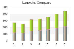
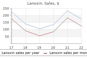
Order discount lanoxin line. iHealth BT Automatic Blood Pressure Wrist Monitor.
The salvage rate was 69% and the overall authors decision to reexplore a failing free flap success rate was 98% arteria zigomatico orbital generic lanoxin 0.25mg on-line. The device is commonly used and come in several shapes and sizes or are cusin anesthesiology and was evaluated by Lindsey et tom-made to fit individual needs arterial hypertension generic lanoxin 0.25 mg line. The expander is al arterial blood pressure buy lanoxin 0.25 mg,356 who demonstrated its usefulness but recomusually connected to a subcutaneous valve through mended further study to develop specific guidewhich isotonic saline is injected for incremental lines before it was universally accepted in clinical expansion hypertension genetics buy cheapest lanoxin. The interval ranges from 3 to delineate the difference between arterial blood pressure chart daily buy 0.25 mg lanoxin with amex, venous blood pressure medication kidney cancer buy genuine lanoxin, 10 days. Comparing tially reserved for experimental puposes only, and simultaneous laser Doppler flowmetry and transcuwill not be discussed here. Although rapid expansion is posclosure by creating, stretching, and approximating sible in some circumstances, preserving tissue integskin flaps. Van Rappard et al10 evaluated the differences in clinical use of controlled skin expansion. He placed a rubber balloon subcutaneously beneath the temsurface area of expanded tissue in relation to shape poral scalp and postauricular skin. For expanders with a months the balloon was gradually expanded, increasround base, a rectangular base, or a crescentic base, ing the skin area by approximately 50% or enough to the respective gains were 25%, 38%, and 32%. Muscle and fat both inflating expander containing hypertonic sodium diminish in mass in response to expansion, and chloride crystals within a shell that gradually fills while there is no loss of muscle function, the loss of through osmosis. The ening of the epidermis after 5 weeks of expansion, concept of a self-inflating expander was explored as well as significant thinning of the dermis and further by Wiese,13 who incorporated a copolymer 20 subcutaneous tissues. Leighton and associates of methylmethacrylate and N-vinyl-2-pyrrolidone found differential thinning of all tissue layers except in a gel casing capable of generating a maximum the epidermis, which was unchanged. The author notes that this Johansson21 also report significant increase in epiexpander is biocompatible and holds promise in dermal thickness, but 6 months after the end of the area of tissue expansion without the disadvanexpansion the epidermis had returned to normal tages noted above. Olenius, Dalsgaard, and Wickman22 studBerge and colleagues14 reported direct closure ied the mitotic activity of human skin samples after with Hydrogel tissue expanders in 9 of 10 patients. This confirmed earlier findings of increased mitosis17 and suggested a net gain of tisthe expanders was 10 mm, and over 20 days they were inflated to 100 mm. Austad and colleagues16,17 nism by which strain causes an enhancement of celstudied changes in the epidermis, dermis, and sublular growth appears to be a network of several intecutaneous tissue of expanded guinea pig skin. Comgrated cascades, implicating growth factors, cytospared with normal skin, expanded skin showed a keleton, and the protein kinase family. Their concluNormal human skin is continually undergoing sions signified that skin expansion is not simply a stretching and relaxation. Collagen fibers exist in a matter of stretching skin but the actual formation of convoluted form and, unlike fibers made of elastin, additional new skin with all the attributes of the are incapable of returning to their relaxed state original tissue. If the limits of the elastic Argenta and coworkers18 summarize the fibers are exceeded, permanent deformation of histomorphologic changes occurring in expanded collagen may result. The epidermis does not change in thickness, changes in the orientation of collagen fibers in the although there is an undulation of the basal lamina dermis as a result of skin stretching. The surrounding utes of stretching with a skin-stretching device, the dermis decreases in thickness considerably, and fibers became aligned in the direction of the stretchincreased numbers of fibroblasts and myofibroblasts ing force, perpendicular to the wound margin. The fibrous dynamic realignment of collagen fibers explains the capsule that forms around the implant consists of significantly decreased wound closing tension thick bundles of collagen fibers and elongated resulting from skin stretching and explains how skin fibroblasts and myofibroblasts. This effect was more pronounced fragmentation of elastic fibers, while the hypoderafter 5 weeks than in the first 1 or 2 weeks of mis, which was in contact with the expander capexpansion. Lee, Squier, and Bardach27 had previsule, did not manifest accentuated fibrosis. The underlying pectoralis major muscle xyproline and a net accumulation of collagen in showed considerable ultrastructural damage under expanded skin compared with normal skin. Kernahan, and Bauer29 likewise noted increased Tissue expansion of irradiated pig skin shows no total collagen content in expanded skin, which further histopathological changes beyond those resulted in a theoretical net gain in the dermal layer caused by irradiation and is indistinguishable from as well as in the epidermal layer. Knight and 30 nonexpanded irradiated skin in the porcine coworkers confirmed increased collagen content 37,38 model. Radiation did reduce the overall area of in expanded dermis of pigs, and speculated that it 38 expanded skin by 23% in one study. Working on could be due to tensile factors during expansion 39 a rabbit model, Goodman and associates note which stimulated biosynthetic activity or mitosis of increased epidermal thickness but no dermal or fibroblasts. The mechanism of action was thought the histologic changes evident in expanded skin to be the normal process of wound healing in addilend support to the concept that skin expansion is a tion to a delay phenomenon. Cherry, Pasyk and others40 comand Jurell report adrenergic supersensitivity in expanded pig skin, suggesting sympathetic denerpared the survival of expanded and delayed flaps vation as a result of expansion. Pasyk, Austad, and Cherry33 note formation of a Expanded flaps showed a 117% increase in survivcapsule around every silicone expander. Specimen angiograms of tains fibrin-like filaments and a cellular layer with expanded skin showed evidence of increased vasmacrophages. Conrecommended including the expander capsule in tains elongated fibroblasts and myofibroblasts orithe flap at the time of transfer for its contribution to ented parallel to the surface of the implant. Has established vessels loosely interspersed with collagen the viability and capillary blood flow of expanded fibers. Compared with acutely Once an expander is removed, the surrounding raised random-pattern flaps, both expanded and fibrous capsule rapidly thins. Matturri and colleagues34 delayed skin exhibited increased total capillary biopsied previously expanded skin at least 1 year blood flow, and this increase paralleled flap surafter expander removal and found normal-appearing vival. The survival length of random flaps in skin epidermis with normal mitotic activity. The dermis overlying tissue expanders was also increased, showed only minimal degree of elastosis and zonal whether the expander was inflated or not. It appears that cyclic loading is the most effecexpansion on ischemia in free flaps. Skin creep the control and sham groups, preexpanded skin alone does not account for all the extra skin during flaps demonstrated a statistically significant increase serial expansion, and factors such as recruitment, (700%) in perfusion as measured by fluorescein. Austad documented a true tisinflation over many weeks and months; the risk of sue dividend from expansion that was thought to infection and implant exposure from the protracted result from the increased mitotic activity of the stressed 44 presence of the expander, particularly in poorly tissues. Vander Kolk and others reported a 32% vascularized areas; and the cosmetic and functional increase in midhorizontal length and 44% increase deformities of buried expanders and valves. After states that these shortcomings of slow expansion flap elevation and inset, the overall increase in surare eliminated by the intraoperative expansion techface area available for coverage was 30%. Instead, they attribute the mechanism of mechanical stretching changes the elasticity and alignexpansion to another, undefined form of subcutament of collagen by a process called creep. Shapiro48 combines acute cycled expansions with the time-dependent plastic deformation of any material or tissue in response to constant stress. Nevertheless, the author states that unto a given constant length, the force required to dermining must still be considered the most impormaintain it is gradually decreased. To date expanders have been used to good effect in the head and neck, the extremities, the trunk, and for breast reconstruction. Expanders are generally contraindicated in areas of poorly vascularized tissue, where there is localized infection, or if there is a higher-thanaverage risk of recurrent cancer. Joss and coworkers54 note that advancement flap Wee, Logan, and Mustoe49 describe a continureconstruction wastes tissue (in dog-ears) at either ous infusion device that maintains a constant end of the defect, and instead recommend transexpander pressure and shortens the time to full posing the expanded tissue into the wound bed expansion by two-thirds. The expander can be of any the efficacy of continuous versus intraoperative tisshape but should be twice as wide as the defect to sue expansion in a pig model, and find three times be covered. He reminds us properties of the skin during rapid and slow tissue that tissue expansion results in a distortion of body expansion for breast reconstruction. Elasticity did not change significantly and neither did hysteresis (a measure of Potential complications of tissue expansion include the skin turgor and plasticity). In summary, there infection, hematoma, seroma, expander extrusion, were minimal differences in skin properties between implant failure, skin necrosis, pain, and neurapraxia. Austad56 notes a remarkgested to relieve pain during expansion,61 but Sinow able absence of disasters in a survey of more than and Cunningham62 report no difference in pain 50,000 tissue expansion procedures, and points out after expansion between patients receiving that the overall incidence of complications associlidocaine analgesia and placebo. Only recounts four cases of partial flap necrosis after the by adding sodium bicarbonate to commercially expander had been removed and the flap advanced available lidocaine to raise its pH to 8. Minor complications were noted in 17% and tion that could lead to lidocaine overdose in the included pain on expansion, seroma, and widening event of implant failure. Argenta and associates18 also noted a 24% Infrequent reports of erosion and deformation of complication rate early in their series, but this subbone underlying an expander have appeared in sequently fell to 7%. Infection is usually reported in the literature, specifically rib concavity with tho1% of cases, and only in patients with predisposing racic skin expansion and calvarial deformity and factors. The most frequent cause of exposure is an remodeling with scalp expansion in children. Sharp edges in the scalp of a child caused by erosion of the or irregular folds in the prosthesis should also be outer table of the skull and bone spur formation smoothed out or risk thinning of the shell from fricfrom pressure by the expander. Argenta recommends waiting for 2 weeks after implantation of the expander before beginning inflation. Plast Reconstr Congress of the International Society of Reconstructive MiSurg 10:149, 1952. Koshima I, Inagawa K, Urushibara K, Moriguchi T: Paraumand clinical implications. Plast Reconstr Surg 102:599, bilical perforator flap without deep inferior epigastric 1998. Koshima I, Inagawa K, Yamamoto M, Moriguchi T: New the head and neck: anatomic study and clinical applicamicrosurgical breast reconstruction using free paraumbilical tions. Koshima I, Moriguchi T, Fukuda H, et al: Free, thinned, venous territories (venosomes) of the human body: exparaumbilical perforator-based flaps. Plast Reconstr microvascular anastomoses: an experimental study and Surg 109:2197, 2002. Nakajima H, Imanishi N, Fukuzumi S, et al: Accompanyconsensus on perforator flap terminology: preliminary ing arteries of the lesser saphenous vein and sural nerve: definitions. Cho B-C, Lee J-H, Byun J-S, Baik B-S: Clinical applications Guide to Clinical Practice, 2nd Ed. In: Cohen M (ed), Mastery of Plastic eral thigh flaps for reconstruction of head and neck and Reconstructive Surgery. Wei F-C, Jain V, Suominen S, Chen H-C: Confusion comprehensive classification of V-Y plasty and its anaamong perforator flaps: what is a true perforator flap Plast logues: the pros and cons of inverted versus ordinary Reconstr Surg 107:874, 2001. Cervical and Clavicular Tubed retrospective comparison of abdominal muscle strength Skin flaps. Chen H-C, Tang Y-B: Anterolateral thigh flap: an ideal soft fasciocutaneous flaps. Kimata Y, Uchiyama K, Ebihara S, et al: Anatomic variaof the septocutaneous vessels of the leg. Plast Reconstr Surg tions and technical problems of the anterolateral thigh 76:354, 1985. Hsieh C-H, Yang C-C, Kuo Y-R, et al: Free anterolateral Gegenbaurs Morphol Jahrb (Leipzig) 121:492, 1975. Plast Reconstr Surg supplied by the vascular axis of the sensitive superficial 107:1766, 2001. Koshima I, Inagawa K, Urushibara K, et al: Deep inferior Plast Reconstr Surg 89:1115, 1992. Nakajima H, Imanishi N, Fukuzumi S, et al: Accompanytion of craniofacial contour deformities. Plast Reconstr ing arteries of the cutaneous veins and cutaneous nerves Surg 106:10, 2000. Marchetti C, Gessaroli M, Cipriani R, et al: Use of venoadipofascial and/or neuroadipofascial pedicled perforator flaps in skull base reconstruction after tumor fasciocutaneous flap. Ann Plast Surg neuroadipofascial pedicled fasciocutaneous flap: a radio50:90, 2003. Fraccalvieri M, Verna G, Dolcet M, et al: the distally based breast reconstruction with the deep inferior epigastric superficial sural flap: our experience in reconstructing the perforator flap. Plast Reconstr Surg flap: clinical experience and evolution to the posterior 103:1191, 1999. Celik N, Wei F-C, Lin C-H, et al: Technique and strategy Reconstr Surg 111:837, 2003. Chen S-L, Chen T-M, Chou T-D, et al: the distally based analysis of 15 complete and partial failures in 439 cases. Wungcharoen B, Pradidarcheep W, Santidhananon Y, on blood flow and metabolism in a skin flap. Plast Reconstr Chongchet V: Pre-arterialisation of the arterialised venous Surg 79:375, 1987. Wungcharoen B, Santidhananon Y, Chongchet V: PrePlast Reconstr Surg 75:88, 1985.