Quinn Capers, IV, MD, FACC, FSCAI,
- Associate Dean for Admissions
- The Ohio State University College of Medicine
- Director, Peripheral Vascular Interventions
- Assistant Professor of Internal Medicine
- The Ohio State University Medical Center
Once the foreign body is identified virus 48 hours to pay fine discount 500mg cephalexin free shipping, removal should be addressed at the time of presentation infection after knee replacement cheap cephalexin 250mg on-line. The surgeon should also communicate before the patient enters the operating suite antibiotic resistance powerpoint discount cephalexin 500mg mastercard. The operwith the anesthesiologist to confirm the depth of anesating surgeon should be gloved and in position before thesia so that laryngospasm is avoided upon withdrawal induction antibiotic induced diarrhea treatment discount 500 mg cephalexin mastercard, and the plan for induction should have of the bronchoscope treatment for dogs eye discharge cheap 250 mg cephalexin overnight delivery. The bronchoscope should be already been discussed between the surgeon and the advanced again to rule out further foreign bodies in the anesthesiologist antibiotic resistance prevalence discount cephalexin 500 mg. If an esophageal foreign body has been diagnosed or is Nuts and other foods may require multiple passes. The esophagoscope may be introduced with the Flexible suction catheters can be advanced down the help of a laryngoscope or under direct vision. Once the side port to remove secretions and facilitate visualizaforeign body has been identified, extraction may require tion. Depending on the ease of extraction, the child removing the entire telescopic forceps and the esophagomay require a postoperative chest x-ray and close folscope complex. Care should be taken to avoid accidental low-up to rule out the development of pneumonia. At least one more pass of the esophagoscope should be performed to check for multiple foreign bodies or Most children make a full recovery without permamucosal damage. The esophagoscope should never be nent sequelae from aerodigestive tract foreign body forced, but should be gently advanced, taking care to have ingestion. Delays in the diagnosis cause the most severe the lumen centered in the field of vision. Children who have a delayed or technically the distance from the esophageal inlet to any signs of difficult extraction should be observed postoperatively mucosal damage should be recorded. A randomized clinical trial of the management of esophmost useful predictors of complications. The cricoid cartilage develops abnormally and may be elliptical or flattened in shape, causing cartilaginous stenosis. The remainder of subglottic stenosis is the following tests are diagnostic: considered to be iatrogenic; airway instrumentation with Flexible laryngoscopy both tube size relative to the airway and the duration Posteroanterior and lateral neck and chest x-rays of intubation plays a role. Rigid endoscopy and microlaryngoscopy Acquired subglottic stenosis more often involves soft tissue stenosis in contrast to the congenital form, which General Considerations results in cartilaginous stenosis. The characterization of stenosis during diagnostic advances in endotracheal tube and ventilation manageendoscopy, including the location, severity, and length of ment in the last 30 years have decreased the incidence of the stenosis, is extremely important and helps to direct subglottic stenosis in the neonatal population to < 1%. It is these patients who provide some of the greatest diagnostic Laryngomalacia is the most common cause of neonatal and management challenges for the otolaryngologist. Laryngomalacia is generally sis, and supraglottic and glottic stenosis, have prompted classified into three main types based on the anatomic otolaryngologists to continue to refine surgical airway portion of the supraglottic structures that is prolapsing, reconstruction techniques. The problem of pediatric laryngotracheal stenosis: a clinposes that immature cartilage lacks the stiff structure of ical and experimental study on the efficacy of autogenous cartimore mature cartilage. The second theory suggests laginous grafts placed between the vertically divided halves of the posterior lamina of the cricoid cartilage. Laryngomalacia can be exacerbated by other entihistory of subglottic stenosis diagnosis and management. Reflux in infants with laryngomalacia: results of 24-hour double-probe pH monitoring. Circumferential acquired subglottic 59% had undergone laryngotracheal reconstruction, and stenosis. This condition may be congenital or secondary to an abnormality along the course of the recurPrevention rent laryngeal nerve. The most common etiology is secondary to hydrocephalus from a malformation such as Advances in airway management of the premature infant Arnold-Chiari. Once the primary cause has been addressed, over the last 30 years have brought incidence rates of subthe paralysis should resolve. Glottic stenosis is generally iatrogenic, resulting from either traumatic intubation involving a similar pathogeneClinical Findings sis as subglottic stenosis or prior laser procedures on the A. The etiology of supraglottic stenosis may also involve prior airStridor is one of the foremost features of airway patholway laser surgery or previous open airway procedures ogy. A more typical presentation of acquired subglottic involving long-term indwelling stents with subsequent stenosis, however, may be of a premature infant with a granulation tissue and fibrosis formation. The pathogenesis of acquired laryngeal web generally involves development of an inflammatory process in reaction to the initial insult, with subsequent maturation and scar formation. Airway fluoroscopy may scale, although still somewhat subjective, is an attempt to demonstrate coexisting pathology, such as tracheomalaprovide an objective parameter of stenosis severity. Other cia, but is dependent on the expertise of the radiologist important characteristics to consider in the preoperative for the diagnosis. Preoperative barium swallow is recomendoscopic exam include the length of the stenosis, how mended if a child has a history of feeding difficulties. This test have evaluated only postoperative voice quality after airway involves a probe in the pharynx (above the upper esophreconstruction surgery, ideally, a preoperative exam would ageal sphincter) and in the esophagus (above the lower add value for comparison and, in the future, may be esophageal sphincter) for detection of acid over a 24-hour included more routinely in the preoperative evaluation. Gastroesophageal reflux: a critical factor in pediatric subrepeat pH probe is still found to be positive, antireflux surglottic stenosis. Voice problems after pedition, premature spillage, aspiration, hypopharyngeal clearatric laryngotracheal reconstruction: videolaryngostroboance, hypopharyngeal pooling, or laryngeal and hypophascopic, acoustic, and perceptual assessment. This planned alteration resulted in G-tube placeties in all patients postoperatively. Benefit of feeding assessment before pediatric reconstruction was modified with the goal of preventing airway reconstruction. It is also important to consider the possibiling the stent position can sometimes resolve symptoms. Diagnoses to consider Some children may require feeding tube placement include laryngomalacia; vocal fold paralysis; laryngeal until stent removal to allow for adequate nutritional web, cyst, or cleft; laryngocele; subglottic hemangioma; support. CarComplications can be grouped into three general catetilage grafts can also prolapse into the airway, causing restenosis. These complications may be avoided if rougories: intraoperative, early postoperative, and late posttine postoperative endoscopy is performed at regular operative. Posterior glottic stenosis may require expansion surgery with a posterior cartilage Intraoperative complications can include either pneugraft. Extreme care should be taken when dissecting rior commissure asymmetry, formation of a glottic web, around the pleura or recurrent laryngeal nerve to avoid or vocal fold scarring. Seroma or cutaneous fistula is another potential complication after wound infection can also occur. Some surgeons recoma long-standing tracheotomy and may require repair mend empiric or culture-directed antibiotic therapy durwith excision of the epithelial-lined tract. The Arytenoid prolapse may occur, most commonly after surgeon and intensive care unit staff must balance the cricotracheal resection or extensive laryngotracheal risk of stent dislodgement with the risks of sedation and expansion procedures. Postoperative cians have found alternative airway management methsupraglottic collapse can be an extremely challenging ods to avoid reinstrumentation of the airway (eg, the use problem. Revision airway surgery may increase longed nasotracheal intubation can also cause alar necrodecannulation rates. Noninvasive positive-pressure ventilation facilitates tracheal extubation after laryngotracheal specific procedure appropriate for that patient. Complications of pediatric laryngotracheal reconstruction: prevention strategies. Single-stage reconstruction gastroesophageal reflux disease as indicators of restenosis. Leak pressures of mild to moderate lesions, and (3) airway expansion sur< 20 cm H2O are associated with successful extubation. These procedures involve cartilagment option as temporary, with the eventual goal being inous grafting and indwelling stents, but the tracheotomy decannulation either with or without airway expansion is retained and not removed until after stent removal. Tracheotomy is not without complications, howsion of the lateral cricoid walls without graft placement ever, and requires a significant amount of education about may allow for even greater expansion of the subglottic and resources for postoperative care, which can sometimes lumen. The initial concern about using at least 1500 g without significant confounding cardiac cartilage grafts was that they may not survive, but histoor pulmonary compromise and evidence of anterior sublogic studies have demonstrated excellent survival and glottic stenosis on bronchoscopy. Important graft properties include that anterior cricoid cartilage is divided in the midline, and (1) it is of the correct depth so as not to protrude into the the incision is extended superiorly through the lower airway, (2) the perichondrium is left intact and faces the third of the thyroid cartilage and inferiorly through the lumen, and (3) the graft is adequately secured. The classic anterior costal carti1 mg/kg/d 24 hours before extubation and for 5 days lage graft is shaped like a boat with flanges, which, when postoperatively. The intended outcome is for a fibrous the graft is inserted into the anterior cricoid incision, are band to form at the incision site, causing the airway to flush with the lateral native cricoid ring. The success rate taken that the graft does not protrude into the lumen, of the procedure is reported to be between 70% and thereby compromising the lumen diameter. There has been concern that the procedure may niques have now been described for endoscopic placedisrupt future cartilage growth, but this concern has not ment of cartilage grafts to address the posterior compobeen demonstrated thus far. Long-term stenting can be associated with dure, with a duration ranging from 1 week to 3 significant complications. For multistage procedures, stents have been kept in place from several weeks to over a year. Rarely, a child with signifiideally, technically adequate expansion surgery should cant cardiac or pulmonary compromise or failure to not require long-term stenting. Tracheotomy is the tions that may impact healing, such as diabetes and procedure of choice for severe laryngomalacia. Suprachronic steroid dependence, should also be considered glottoplasty may be used in less severe cases. A wide-mouthed laryngoscope (eg, a extensive procedures designed to remove rather than Lindholm laryngoscope) may be helpful in providing expand the stenotic segment have also been developed, the best view of the supraglottis. The procedure is usuincluding cricotracheal resection, slide tracheoplasty, and ally performed under general anesthesia with spontaneeven tracheal homograft transplantation. Care must be taken to is now being implemented as a first-line treatment for avoid lasing adjacent surfaces to prevent scar formation; some patients with severe and even moderate stenoses. Most patients can be cricoid arch with preservation of a posterior mucosal extubated at the end of the procedure, and often a short flap along the posterior cricoid plate. Involvement of the vocal folds is a contraindication, and generally, a superior Posterior glottic stenosis and bilateral vocal fold paralysis margin of 3 mm is recommended for success. Inferior may often be difficult to distinguish and can have similar resection margins have extended as low as the second presenting symptoms. Tracheotomy is an option for success and a suprahyoid release has been used to both diagnoses, but is generally not considered an optimal achieve this. A subperichondrial traor complete arytenoidectomy, cordotomy, and partial corcheal dissection is recommended to avoid nerve injury.
Haematoma forms due to bleeding from torn blood vessels antimicrobial towels buy cephalexin 500mg free shipping, flling the area surrounding the fracture infection 7 weeks after dc purchase 500 mg cephalexin with visa. Local infammatory response occurs at the site of injury with exudation of fbrin antibiotics h pylori discount cephalexin 250 mg, polymorphs and macrophages bacterial zoonoses discount 500 mg cephalexin visa. Ingrowth of granulation tissue begins with neovascularisation and proliferation of mesenchymal cells from periosteum and endosteum bacteria 3 shapes purchase cephalexin master card. The cells of inner layer of the periosteum have osteogenic potential and lay down collagen as well as osteoid matrix in the granulation 97 tissue infection control risk assessment cheap generic cephalexin uk. The woven bone is cleared away by incoming osteoclasts and the calcifed cartilage disintegrates. Fibrous union may result instead of osseous union if the immobilisation of fractured bone is not done. Non-union may result if some soft tissue is interposed between the fractured ends. Delayed union may occur from causes of delayed wound healing in general such as infection, inadequate blood supply, poor nutrition, movement and old age. These fbrils grow along the track of degenerated nerve so that in about 6-7 weeks, the peripheral stump consists of tube flled with elongated Schwann cells. On injury, the cut ends of muscle fbres retract but are held together by stromal connective tissue. However, in large destructive lesions, the smooth muscle is replaced by permanent scar tissue. This occurs by proliferation from margins, migration, multilayering and differentiation of epithelial cells. However, in parenchymal cell damage with intact basement membrane or intact supporting stromal tissue, regeneration may occur. Stem cells are the primitive cells which have 2 main properties: i) They have capacity for self renewal. Stem cells exist in both embryos and in adult tissues: fi In embryos, they function to generate new organs and tissues; their presence for organogenesis has been an established fact. For example, stem cells in the bone marrow which sponateously differentiate into mature haematopoietic cells has been known for a long time. Some of the major clinical trials on applications of stem cells underway are in the following directions: 1. Bone marrow stem cells Haematopoieitc stem cells, marrow stromal cells and stem cells sourced from umbilical cord blood have been used for treatment of various forms of blood cancers and other blood disorders for about three decades. Neuron stem cells these cells are capable of generating neurons, astrocytes and oligodendroglial cells. Islet cell stem cells Clinical trials are under way for use of adult mesenchymal stem cells for islet cells in type 1 diabetes. Cardiac stem cells It is now known that the heart has cardiac stem cells which have capacity to repair myocardium after infarction. Skeletal muscle stem cells Although skeletal muscle cells do not divide when injured, stem cells of muscle have capacity to regenerate. Adult eye stem cells the cornea of the eye contains stem cells in the region of limbus. These limbal stem cells have a potential therapeutic use in corneal opacities and damage to the conjunctiva. Skin stem cells In the skin, the stem cells are located in the region of hair follicle and sebaceous glands. Which of the following type of leprosy is not included in RidleyJopling classifcationfi They do not have capacity to multiply in response to stimuli throughout adult life 21. Connective tissue in scar is formed by the following types of fbrillar collagen: A. Following is the range of host-organism inter-relationship, which may vary quite widely: 1. Prions are transmissible agents similar to infectious particles but lack nucleic acid. However, if the host is old, debilitated, malnourished, or immunosuppressed due any etiology, he is susceptible to have manifestations of infection. However, confrmation in most cases requires either application of special staining techniques or is confrmed by molecular biologic methods as under: 1. Direct identifcation of the organism in tissues is possible by fuorescence antisera methods. Plague is a zoonotic disease and spreads by rodents, primarily by rats, both wild and domestic; others being squirrels and rabbits. It is a spore-forming bacillus and the spores so formed outside the body are quite resistant. The disease occurs as an exogenous infection by contact with soil or animal products contaminated with spores. Depending upon the portal of entry, three types of human anthrax is known to occur: i) Cutaneous form by direct contact with skin and is most common. The organisms proliferate here and stimulate the bronchial epithelium to produce abundant tenacious mucus. Within 7-10 days after exposure, catarrhal stage begins which is the most infectious stage. The infection is transmitted through vaginal or anal intercourse and by autoinoculation. The dermis and subcutaneous tissues are infltrated by numerous histiocytes containing many bacteria called Donovan bodies, and lymphocytes, plasma cells and neutrophils. Chlamydia are no more considered as flterable viruses as was previously thought but are instead intracellular gram-negative bacteria. M/E the lymph nodes have characteristic stellate-shaped abscesses surrounded by a zone of epithelioid cells (granuloma). Healing stage of the acute lesion takes place by fbrosis and permanent destruction of lymphoid structure. There is regional nodal enlargement which appears about 2 weeks after cat-scratch, and sometimes after thorn injury. M/E Main features are as under: i) Initially, there is formation of non-caseating sarcoid-like granulomas. The following groups and subtypes of streptococci have been identifed and implicated in different streptococcal diseases: 1. Group A or Streptococcus pyogenes, also called b-haemolytic streptococci, are involved in causing upper respiratory tract infection and cutaneous infections (erysipelas). Group D or Streptococcus faecalis,also called entero cocci are important in causation of urinary tract infection, bacterial endocarditis, septicaemia etc. Untypable a-haemolytic streptococci such as Streptococcus viridans constitute the normal fora of the mouth and may cause bacterial endocarditis. Pneumococci or Streptococcus pneumoniae are etiologic agents for bacterial pneumonias, meningitis and septicaemia. On degeneration of these microorganisms, the plasmids are liberated which produce many toxins responsible for the following clostridial diseases depending upon the species: 1. The material discharged from the sinuses is in the form of grains consisting of colonies of fungi or bacteria. Mycetomas are of 2 main types: fi Mycetoma caused by actinomyces (higher bacteria) also called actinomycetoma comprises about 60% of cases. The lesions extend deeply into the subcutaneous tissues, along the fascia and eventually invade the bones. M/E They drain through sinus tracts which discharge purulent material and black grains. In human beings, Candida species are present as normal fora of the skin and mucocutaneous areas, intestines and vagina. The organism becomes pathogenic when the balance between the host and the organism is disturbed. These superfcial fungi are spread by direct contact or by fomites and infect tissues such as the skin, hair and nails. Viruses causing haemorrhagic fevers were earlier called arthropod-borne (or arbo) viruses since their transmission to humans was considered to be from arthropods. However, now it is known that all such viruses are not transmitted by arthropod vectors alone and hence now such haemorrhagic fevers are classifed according to the routes of transmission and other epidemiologic features into 4 groups: 1. Of these, mosquito-borne viral haemorrhagic fevers in which Aedes aegypti mosquitoes are vectors, are the most common problem the world over, especially in developing countries. Two important examples of Aedes mosquito-borne viral haemorrhagic fevers are yellow fever and dengue fever. Chikungunya virus infection is primarily a disease in nonhuman primates but the infection is transmitted to humans by A. The disease is endemic in parts of Africa and Asia and occurs sporadically elsewhere. Its general clinical features range from a mild afebrile illness similar to common cold by appearance of sudden fever, headache, myalgia, malaise, chills and respiratory tract manifestations such as cough, soar throat to a more severe form of acute respiratory illness and lymphadenopathy. Depending upon its antigenic characteristics of the nucleoprotein and matrix, 3 distinct types are known: A, B and C. Out of these, infuenza type A is responsible for most serious and severe forms of outbreaks in human beings while types B and C cause a milder form of illness. The virus is transmitted into the human body by the bite of infected carnivores. The organism enters a peripheral nerve and then travels to the spinal cord and brain. Parasites may cause disease due to their presence in the lumen of the intestine, due to infltration into the blood stream, or due to their presence 107 inside the cells. The condition is particularly more common in tropical and subtropical areas with poor sanitation. The parasite occurs in 2 forms: fi a trophozoite form which is active adult form seen in the tissues and diarrhoeal stools; and fi a cystic form seen in formed stools but not in the tissues. M/E the lesions of amoebiasis include amoebic colitis, amoeboma, amoebic liver abscess and spread of lesions to other sites. While Plasmodium falciparum causes malignant malaria, the other three species produce benign form of illness. The disease is endemic in several parts of the world, especially in tropical Africa, parts of South and Central America, India and South-East Asia. The main clinical features of malaria are cyclic peaks of high fever accompanied by chills, anaemia and splenomegaly. Major complications occur in severe falciparum malaria which may have manifestations of cerebral malaria (coma), hypoglycaemia, renal impairment, severe anaemia, haemoglobinuria, jaundice, pulmonary oedema, and acidosis followed by congestive heart failure and hypotensive shock. The lymphatic vessels inhabit the adult worm, especially in the lymph nodes, testis and epididymis. The eggs are passed in human faeces which are ingested by pigs or they infect vegetables. These eggs then develop into larval stages in the host, spread by blood to any site in the body and form cystic larvae termed cysticercus cellulosae. M/E the cysticercus may be single or there may be multiple cysticerci in the different tissues of the body. The cysts may occur virtually anywhere in body and accordingly produce symptoms; most common sites are the brain, skeletal muscle and skin. Cysticercus consists of a round to oval white cyst, about 1 cm in diameter, contains milky fuid and invaginated scolex with birefringent hooklets. The infection may be acquired by the foetus during intrauterine life, or perinatally and damage the foetus or infant. Dengue haemorrhagic fever is characterised by following laboratory fndings except: A. Granuloma inguinale and lymphogranuloma venereum are similar in following aspects, except: A. The tumours derive their nomenclature on the basis of the parenchymal component comprising them. Malignant tumours of epithelial origin are called carcinomas, while malignant mesenchymal tumours are named sarcomas (sarcos = feshy). However, some cancers are compo sed of highly undifferentiated cells and are referred to as undifferentiated malignant tumours. Although, this broad genera lisation regarding nomenclature of tumours usually holds true in majority of instances, some examples contrary to this concept are: melanoma for carcinoma of the melanocytes, hepatoma for carcinoma of the hepatocytes, lymphoma for malignant tumour of the lymphoid tissue, and seminoma for malignant tumour of the testis. Mixed tumours A few examples are as under: i) Adenosquamous carcinoma is the combination of adenocarcinoma and squamous cell carcinoma in the endometrium.
It is a relatively uncommon lesion as occasionally painful bacteria yogurt lab cheap cephalexin, nonpulsatile swellings over the jaw treatment for sinus infection headache generic cephalexin 500mg amex. The differential diagnosis should include ameloblastomas infection nail bed cheap cephalexin 500 mg without prescription, developmental odontogethe pathogenesis of this lesion is unknown; theories sugnic cysts infection 2 bio war simulation purchase cephalexin visa, central giant cell granulomas virus with diarrhea order cephalexin 500 mg with amex, and central vascular gest that its pathology results from a traumatic episode that lesions antibiotics for diphtheroids uti buy generic cephalexin 500 mg line. Complications include a destructive osteolytic procauses a hematoma to form within the intramedullary cess of the involved jaw. The prognosis is generally good, provided producing osteolysis and an empty bone cavity. The differential diagnosis includes odontogenic keratocysts, central giant cell granulomas, or odontogenic tumors. These cysts Complications include local bone destruction and the are also known as Stafne bone cysts, lingual mandibular displacement of tooth roots. It is believed to be developmental in nature but does not appear at birth and is not Treatment seen in children. Most cases are seen in middle-aged or Surgical exploration is the treatment modality most older adults. Eighty to ninety percent of these defects commonly used to rule out the existence of other more are seen in males. Surgical exploration is not indicated, but these defects contain salivary gland or adipose tissue from the floor of the mouth. There has been a report of a salivary gland neoplasm developing in the lingual mandibular salivary gland depression. A static bone cyst does not require biopsy or excision unless a mass can be identified or imaged or there are clinical findings. These cystic lesions are not classically described in discussions of cystic lesions of the jaw, but, because of their presentation, they may be confused with parotid tumors. There are two types of ganglion cysts: (1) those with walls that consist of fibrous connective tissue and (2) those with walls that are lined by synovial cells. The surgical removal with histopathologic examination of the excised tissue is the treatment of choice for jaw cysts in most cases. X-ray of a cadaveric mandible with a dibular joint should be considered in the differential diagnostatic bone cyst. There is no evidence of a higher incidence which can be acute, recurrent, or chronic. The association drops off dramatically for both men and women after between wear facets, bruxism, and severity of facial pain in age 55. However, many patients claim muscle and joint (eg, blows to the jaw) and indirect (eg, whiplash injupain after a whiplash injury. Microtrauma is caused by clenching from the strained sternocleidomastoid muscle, which and grinding of the teeth. Stress can be a predisposing often refers pain to the ear, or it may be due to injuries factor owing to the disruption of restorative sleep and to other cervical muscles and ligaments. Controversial Causes Studies reporting on the long-term follow-up of patients with disc displacement show the majority are asymptomA. Bruxism, or grinding the teeth during sleep, has been thought to be a predisposing, precipitating, and perpetClinical Findings uating factor. Self-care should be thoroughly explained to patients in language meaningful to them, and it should be reinforced at each visit. Imaging should be reserved for patients Good posture; avoid head-forward position. Imaging is also warranted in patients who have a sudden change in the bite or asymmetry of the mandible. Temporomandibular disorders are divided into articular Yoga and meditation for stress reduction. Studies have shown that dry severe and does not respond to other treatments, opioid needling works just as well, and the difference between analgesics are often beneficial. Oral splints should be used as an adjunct for pain management rather than a definiPhysical therapy has been shown to be helpful for many tive treatment. Heat and ice Treatment with intraoral splints has been shown to have beneficial effects on reducing pain in some patients. Splints reduce the role of occlusal decreasing hypermobility, strengthening and coordinatfactors, reduce loading on the joints, and have a strong ing muscles, and improving muscle endurance. Splints can reduce tooth damage in can be helpful because it promotes increased blood flow patients who grind their teeth and can increase awarethrough the tissue in addition to inducing muscle relaxness of these detrimental oral habits. The evaluation of patient posture is important, get relief and some experience a worsening of symptoms and patients should be taught proper posture. There are possible complications to wearhead position can exacerbate neck pain and a tense jaw ing splints, such as irreversible changes in occlusion that posture can increase jaw and muscle pain. Therefore, splints should be worn for a short to moderate time period and should be regularly moniUltrasound can provide deep and relaxing heat to muscles tored. Nighttime wear is typical and full-time use is and joints, helping to relieve pain and restore function. Another other situations (such as myofascial pain), acupuncture cannula is then inserted with microtools that allow for may be useful as an adjunct treatment or an acceptable debridement, the removal of adhesions, and biopsies. A number of studies of acupuncture and chronic pain found positive results in 41% of them J. Short-term pain reduction with acupuncture treatwhom an identifiable structural defect can be corrected by ment for chronic orofacial pain patients. Preand postoperative physical therapy is importhe lateral pole of the condyle is evident. No the more invasive open joint procedures, so arthrocentesis radiographic changes are found; however, evidence of and arthroscopy should be considered as a first step. It articulates the accompanies the click, it is most often the result of mandibular condyle and the squamous portion of the inflammation in the joint owing to the condyle pressing temporal bone, with the articular disc of dense fibrous on the retrodiscal tissues, synovitis, or capsulitis. Sometimes patients complain about posterior teeth not Acute disc displacement without reduction (closed meeting on the same side, presumably because of swelllock) is characterized by a marked limitation in opening in the joint. It is also distinguished by a deflection Patients often present with a history of pain in the of the mandible to the affected side on opening. It occurs preauricular region, which is aggravated by chewing or with a sudden onset and can be painless or painful. Pain on palpation over clicking is felt or heard, although the patient usually has a history of clicking at one time. The disc is usually anterior to the condyle and blocks the translation of the condyle, preventing normal opening and causing the mandible to deflect to the affected side. Radiographs can show a decreased joint space that might be an indication of a displaced disc. The disc is anterior to the condyle and is either pushed further anterior on opening or is folded on itself. The that is characterized by deterioration and abrasion of condyle remains positioned anterior to eminence. It is accompanied by remodeling of There can be joint pain at the time of dislocation and the underlying subchondral bone. There is usually a hiswith function, and crepitus is often heard over the tory of a self-reducing dislocation. Joint stiffness, often worse on awakening the condyle can be reduced by manually pushing or at the beginning of a meal, can be a problem, and the mandible both downward and backward into the the patient may have a limited range of motion. This reduction can often be done in the office by graphic evidence of degeneration of the condyles can be placing gloved hands, with the thumbs outside the seen. The long-term prognosis is good because and distracting the mandible in a downward direction, osteoarthritis tends to be self-limiting as the joint placing the condyles back into the fossa. Pain is usually elicited with function, and Fibrous ankylosis is restricted mandibular movement with the patient may experience a limited range of motion. This condition Crepitus can be heard over the affected joint and degenresults from fibrous adhesions that attach the condyle to eration of the condyles may be seen on x-rays. There is a marked limited opening, usually < 20 mm, but the condition is not painful. Radiographs show an absence of condydrosarcomas, fibrosarcomas, and synovial sarcomas. Squamous cell carcinomas of the maxillofaof bone cells, which results in the complete immobility cial region and nasopharyngeal tumors are the tumors of the joint. Pain is elicited on palpation side on opening, and there is a marked limited lateral and with function. There can be an open bite on the movement of the mandible to the contralateral side. This positive finding of tumor with imaging, and both imagcondition is marked by a limited opening (< 25 mm), ing and biopsy help confirm the diagnosis. There is often bleeding in the joint, and sequelae can include adhesions, ankylosis, and joint degeneration. The mandible deflects to the affected side and the fracture is evident on an x-ray. Open joint surgery is required to the muscles of mastication are the masseter, temporareduce the fracture only in rare cases. In addition to neoplasms, which are rarely seen, more common muscle disorders may result in pain, redness, swelling, cramping, and contracture. One percent of malignant breast tumors ing muscle pain, usually of mild to moderate intensity. The pain is aggravated by mandibular function when the muscles of mastication are involved. Patients may also present with a sensation of musMyositis is characterized by moderate to severe pain, cle stiffness or tightness and a sensation of their teeth redness, and swelling associated with tissue injury. Inactivating the trigger points condition can result from direct trauma or infection, with a local anesthetic injection, acupuncture, or a often secondary to oral surgery or an intramuscular vapocoolant spray and muscle stretch often relieves the injection. The pathogenesis is now cle area following injury or infection, and diffuse tenthought to be due to changes in the central nervous sysderness is present over the entire muscle. Treatment outcome of appliance therapy in range of mandibular motion is often present. Elevated temporomandibular disorder patients with myofascial pain serum levels indicative of inflammation, infection, or after 6 and 12 months. It is often accompanied by fatigue, irritable bowel rest as well as with function. Basic drug therapy rests on the muscle undergoes fibrotic changes and becomes hard. Familiarity cleidomastoid muscle medially, the trapezius muscle with these specific areas assists in generating a differential laterally, and the middle third of the clavicle inferiorly. The structures that make up the anterior neck include the larynx, trachea, the differential diagnosis in a patient presenting with a esophagus, thyroid and parathyroid glands, carotid neck mass is broad and extensive. The submental triangle defines a region borthe most important element in the evaluation of a dered by the hyoid bone, the paired anterior bellies of neck mass is the age of the patient. In used in the detection and surveillance of head and neck contrast, a neck mass in an adult over the age of 40 cancer. The probability of a benign neck mass in this Serologic Testing age group is low, particularly in the setting of tobacco or alcohol use. Serologic testing can be used in looking for systemic the duration, growth pattern, and absence or presdiseases. For example, antinuclear antibody may be posence of pain are all critical aspects of the history. A itive in Sjogren syndrome, which can present with region-specific review of systems, such as a change in parotid enlargement and lymphadenopathy. Serologic voice, hoarseness, difficulty with swallowing, and ear testing is also important in the diagnosis of many infecpain are important symptoms to note in addition to tious diseases that may present as a neck mass, includgeneralized complaints such as fever, night sweats, and ing tuberculosis, atypical mycobacteria, mononucleoweight loss.
Polyps are often T2imaging as mucosal thickening efficacy of antibiotics for acne buy generic cephalexin from india, retention cysts virus classification discount 250mg cephalexin with amex, polyps what causes antibiotic resistance yahoo purchase 500mg cephalexin otc, sinus hyperintense and may show diffuse or surface enhancement infection quotient purchase cephalexin 250 mg mastercard. A mucocele develops from sinus ostial obstruction and reOrbital complications of sinusitis include preseptal periorsults in opacification and expansion of the sinus bacteria 4 urinalysis discount cephalexin 500mg with mastercard. Intracranial complications include meningitis antibiotic resistance and infection control journal purchase discount cephalexin, the peripheral enhancement of a mucocele distinguishes it from empyema, abscess, thrombophlebitis, and cavernous sinus thromneoplasm. Osteomyelitis rarely complicates sinonasal tensity may represent chronic inspissated secretions, mycetoinfection but may occur with trauma, surgery, or hematogenous ma. Ear and Temporal Bone Complications are related to bony erosion or deformity that may Otitis Media and Mastoiditis involve the scutum, ossicles, mastoid, tegmen tympani, sigmoid Acute and chronic forms of otitis media characteristically produce sinus plate, facial nerve canal, or lateral semicircular canal. Secondary bacterial infection is comcomplications include meningitis, abscess formation, venous mon. It appears as a nonenhancing soft tissue mass with sharply marteal abscess, and neck abscess (Bezold abscess). Intracranial complications result from bony erosion or septic thromOtitis Externa bophlebitis and include epidural abscess, subdural empyema, Otitis externa is often self-limiting. In immunocompromised pameningitis, cerebritis, cerebellitis, brain abscess (usually in the tients, a severe necrotizing form may develop that extends to the temporal lobe or cerebellum), and dural venous sinus thrombosis. Granulation A simple mastoidectomy results in removal of mastoid air cells tissue may be soft or fibrous, contain cholesterol or hemorrhage, but preservation of the external canal wall and ossicles. Primary acquired cholesteatomas the Neck, Oral Cavity, and Jaw result from eustachian or attic obstruction with tympanic memPharyngeal and Retropharyngeal Infection brane (superior pars fiaccida) retraction. Because of ossicular destruction, only a portion of the right incus remains (short arrow). Complications petrous apex opacification, enhancement, and bony destruction (arrows include airway encroachment, osteomyelitis, sinus or orbital inin A) as well as the right dural, cavernous sinus, and orbit involvement volvement, internal jugular vein thrombosis, carotid artery rup(arrows in B and C). Parapharyngeal or retrophaLymphadenitis is the most common cause of lymphadenopathy ryngeal lymphadenitis may do the same, especially in infants and in childhood. Acute adenitis may be bilateral from the skull base to the mediastinum and contains lymph. Subacute or chronic lymphadenitis the more midline perivertebral space edema that may follow is more typical of mycobacterial infections, cat-scratch disease, vertebral osteomyelitis or epidural abscess. Noninfiammatory adenopathy raises clude thickening of the retropharyngeal soft tissues and anterior suspicion for malignancy. Uniform contrast enhancement is comanteroposterior, rotary, or transverse displacement of C1 on C2, mon with viral processes. Mycobacterial adenitis (tuberculous or nontuberculous) is suggested by a nodal mass with central liquefaction, thick margin enhancement, and extension to the skin. Calcification is common but may also be seen in other granulomatous infections, treated lymphoma, and metastatic disease. Cellulitis refers to diffuse bacterial or viral infiammation with edema, swelling, and fat plane obliteration, but no distinct mass. Extensive soft tissue infiltration of multiple tissue planes, including muscle, suggests the more severe condition of fasciitis. Thyroid Infiammation Suppurative submandibular sialadenitis is usually related to sialoHashimoto thyroiditis is the most common acquired thyroid lithiasis. Acute suppurative thyroiditis with abscess suggests a contrast enhancement of the gland and duct walls with ductal dilcongenital pyriform sinus fistula (see. Complications include obstruction, infection, Acute sialadenitis is usually viral or bacterial. Noncalcified stones may be prior infection, dehydration, trauma, surgery, irradiation, certain diagnosed only with sialography. Imaging may show permeative bone destruction, soft sublingual gland duct and manifests as a unilateral mass in the tissue edema, cellulitis, or abscess. Extension below the mylohyoid sequestrum formation, and bony sclerosis indicate chronicity. Chronic sialadenitis may be idiopathic or result from recurrent bacterial infection, ductal obstruction (especially submandibular Neoplastic Processes gland), granulomatous disease, prior irradiation, or autoimmune disease. Sjogren syndrome is an autoimmune disease that may be limited to the Orbit and Globe salivary or lacrimal glands or may also have systemic involveNeoplastic processes of the orbit and globe include ocular tumors, ment. Pathologically, these may be neoplastic tuberculosis, or sarcoidosis may also cause salivary gland enlargeprocesses of mesenchymal, neural, or malformative origin. Such involvement is to be distinguished malformation, plexiform neurofibroma, and teratoma. Tumors most often arising exOsteomyelitis traconally include dermoid-epidermoid, hemangioma, lymphatic Osteomyelitis of the mandible may result from direct inoculamalformation, plexiform neurofibroma, teratoma, neuroblastoma, tion. Mesenchymal Tumors Rhabdomyosarcoma is the most common malignant tumor of the head and neck region. They may also arise elseNeoplastic Processes where and metastasize to the orbit. These aggressive, invasive neoplasms are usually of the embryonal or alveolar subtype. There may Vascular malformations be intracranial extension and regional or systemic metastases. In LangerRhabdomyosarcoma hans cell histiocytosis, there may be solitary or multiple soft tissue Langerhans cell histiocytosis masses with lytic bony destruction of the orbit, sinuses, cranial Leukemia/lymphoma base, or calvaria (see Chapter 8). There may also be pituitary-hypothalamic inOptic glioma volvement with diabetes insipidus, absence of the posterior piNeuroblastoma tuitary bright spot, and hypothalamic or stalk enhancement (see Esthesioneuroblastoma Chapter 8). Primitive neuroectodermal tumor Leukemic infiltration of the orbit may occur in acute lymphoProgonoma blastic leukemia. Juvenile angiofibroma is an inPapilloma vasive fibrovascular mesenchymal tumor of adolescent males that Carcinoma. Metastatic disease Fibromas are mesenchymal tumors that may be isolated and relatively benign. Calcification occurs in more than 90% of and fibrosarcoma, which are discussed later. Bilateral retiNeuroblastoma is the most common neural tumor to invade noblastoma is usually hereditary and may be associated with a the orbit secondarily. It is usupineoblastoma (trilateral retinoblastoma), additional hypothalamic ally a nodular infiltrating mass causing permeative, blastic, involvement (quadrilateral retinoblastoma), and radiation-induced or spiculated bone destruction. Tumors of childhood arising in the nasal cavity, sinuses, and face may be neoplastic or nonneoplastic. They may be of mesOptic pathway tumors are common tumors of childhood and enchymal, neural, cutaneous or mucosal origin. Solitary intraorregional involvement, including orbital or intracranial, is imporbital lesions are rare and include hamartomas, arachnoidal hytant for treatment. Mesenchymal tumors chiasm and optic tracts range from hamartomas and low-grade are of vascular, soft tissue, reticuloendothelial, osteochondroid, astrocytomas to anaplastic astrocytomas. Neural tumors include those of bined intraorbital, intracanalicular, and intracranial optic pathneuroepithelial, neural crest, and nerve sheath origins. Optic gliomas must be distinguished from plastic lesions of cutaneous or mucosal epithelial origin are perioptic tumors such as a schwannoma, neurofibroma, and rare. Extension often occurs into the sphenoid, lar anomalies and tumors and cysts of dental origin are discussed maxillary, and ethmoid sinuses as well as the orbit, middle cranial elsewhere. Sinus or otomastoid obstruction with mucosal pterygopalatine fossa and sphenopalatine foramen, it manifests edema and retained secretions is common. Preoperative catheter as nasal obstruction, epistaxis, facial swelling, proptosis, otitis angiography and therapeutic embolization often facilitate surgimedia, or headache. Bony exAngiomatous polyp and hemangiopericytoma are very rare in pansion and erosion are common, including widening of the childhood but may be mistaken for angiofibroma. Osseous and Chondroid Tumors the orbit and paranasal sinuses are common sites of origin Osseous and chondroid tumors may arise from the facial bones or of rhabdomyosarcoma (see. Langerhans cell histiocytosis is a reticuloendothelial disorder Osteochondroma is a benign osteocartilaginous exostosis that histologically characterized by tissue infiltration with reticulum may arise from the mandible, maxilla, sphenoid bone, zygoma, or cells, histiocytes, plasmocytes, and leukocytes (see Chapter 8). Multiple lesions occur in familial cases and in Olthe involvement may be isolated (formerly eosinophilic granulier disease. Imaging shows a miniature metaphysis, growth plate, and cartiLymphoma is another common malignant tumor of the head laginous cap that are continuous with the bone of origin. Hodgkin disease often manifests nant degeneration is indicated by a disorganized appearance and as cervical lymphadenopathy and spreads contiguously along involvement of the parent bone. Non-Hodgkin lymphoma is often widespread with Fibrous dysplasia is an idiopathic and benign fibro-osseous noncontiguous nodal involvement. The origin may be in the nadisorder that may be monostotic, polyostotic, or part of the sopharynx, sinuses, adenotonsillar region (Waldeyer ring), or saliMcCune-Albright syndrome. Head and neck lymphomas may be associated with frequently involved, unilaterally or bilaterally. Ossifying fibroma is a circumscribed fibrous neoplasm that progresFibromatous tumors are mesenchymal neoplasms that may be sively ossifies. Cementifying fibroma is another fibro-osseinfiltrating pseudoneoplastic process characterized by fibroelastic ous tumor that is aggressive and tends to recur. Desmoid tumor is a well-differentiated form with no Giant cell tumor, giant cell reparative granuloma, aneurysmal bone tendency to metastasize. In other forms there may be widespread visceral ping pathologic findings, and combined lesions are well known. The juvenile form usually involves musculoskeletal have a bony matrix or calcification, cortical erosion, soft tissue structures but not the viscera. Moderate contrast enhancement isodensity to hypodensity and hypointensity, respectively, in the is common. Fibrosarcoma and Ewing has also been reported with lymphatic malformation, venolymsarcoma produce soft tissue masses and permeative bony destrucphatic malformation, and telangiectatic osteosarcoma. Chondrosarcoma is a malignant bone neoplasm of cartilage oriNeuroblastoma, the most common of these tumors, may arise in or gin that may arise de novo, from an osteochondroma, or followinvolve the skull base, nose, sinuses, or orbit, usually as part of ing radiotherapy. They are characterized by small tumors are locally invasive, destroy bone, and may metastasize. Progonothe chondroid form of chordoma may be indistinguishable from mas are rare retinal anlage tumors, often contain melanin, tend chondrosarcoma on imaging. Osteosarcoma may appear as a soft tissue mass with bony deSchwannomas, neurofibromas, and plexiform neurofibromas rarely struction and spiculated periosteal bone reaction, or as a partially arise in the nasal cavity, paranasal sinuses, or nasopharynx. Nasal papillomas are benign mucosal tumors that often extend into They may be hereditary, familial, multicentric, or hormonally acthe maxillary, ethmoid, sphenoid, or frontal sinuses. Paragangliomas are vascular, but slow-growing, tumors that transformation is extremely rare. Complete surgical excision may be tus, and a red retrotympanic mass are characteristic. Differentiation from carotid adenocarcinoma of the nasal cavity and sinuses are extremely and jugular anomalies is necessary. Angiography and therapeutic occur, along with regional extension, nodal spread, and distant embolization are helpful for surgical management. Extensive local involvement is frequent, along with Ear and Temporal Bone intracranial invasion and metastases. Classically, there is no prior infiammation, trauma, cations include internal jugular vein invasion, compression, and or surgery. Metastasis Temporal bone involvement is uncommon and usually monothe most common metastatic tumors of the temporal bone are stotic in fibrous dysplasia. Exostosis is a common, benign bony hyperplasia of the external Neck, Oral Cavity, and Jaw canal. These are to be distinguished mor that is usually unilateral and more often arises in the outer from lymphadenopathy, cellulitis, and abscess. Primary malignant tumors of the pediatric head and neck vary Of the nerve sheath tumors in the region, neurofibromas. Neuroblastoma usually arises in infants and young chilAcoustic or vestibular schwannoma is rare in childhood, sugdren. Characteristically, there is an enhancand signs may be related to associated lymphadenopathy, paing mass that expands the facial canal, jugular foramen, or hyporanasal sinus or ear involvement, aerodigestive compromise, or glossal canal. Fibromatosis colli (also known as congenital muscular torticollis) dermoid-epidermoid is to be distinguished from cephalocele (see is a common benign condition of the neonate and young infant Chapter 8) and vascular anomalies. Suggested causes inTeratomas arise from pluripotential cells and usually manifest clude in utero deformation and birth trauma. There is venous at birth as large neck masses causing respiratory or swallowing hemorrhage evolving to fibrosis. Treatment usually consists of physical therahas the same echogenicity, density, and intensity, respectively, as py, but surgery may be required.
Code descriptions vary depending on whether a cardiopulmonary bypass (heart-lung) machine was used during the procedure antibiotic beginning with c buy cephalexin 500mg lowest price. Medications or balloon valve angioplasty are the less invasive treatments; however antibiotic impregnated beads purchase cephalexin 500mg mastercard, valve replacement may be required infection xrepresentx lyrics buy discount cephalexin 500 mg on line. The delivery catheter is inserted into an artery using a percutaneous infection eyes purchase line cephalexin, open antibiotics for uti azithromycin best 500mg cephalexin, or transaortic approach infection xenophobia cheapest generic cephalexin uk. Her physician believes that because she is symptomatic, she should have her mitral valve replaced. Bypass is discontinued and the atrium and sternum are closed in the usual fashion. The codes include endarterectomy (removal of the inner lining of an artery) and angioplasty (blood vessel repair). Do not unbundle the codes and report the endarterectomy or angioplasty separately. Also, the procedures often require the use of cardiopulmonary bypass to allow the surgeon to repair the defect while the heart is without blood flow, which makes a difference in the choice of codes. Coronary artery bypass Arteries deliver oxygenated blood to all areas of the body, and veins return the blood that is full of waste products. Sometimes the arteries clog to the point that the heart muscle begins to perform at low levels due to lack of blood (reversible ischemia) or actually die (irreversible ischemia). Reversible ischemia means that if the bloodflow is increased to the heart muscle, the heart muscle may again begin to function at normal or near-normal levels. The diseased portion of the artery is bypassed by attaching a healthy vessel above and below the diseased area and allowing the healthy vessel to then become the conduit of the blood, thus bypassing the blockage. Blockage can also be pushed to the sides of the coronary arterial walls by a procedure in which a balloon is expanded inside the artery. To correctly report coronary bypass grafts, you must know whether an artery (33533-33548), a vein (33510-33516), or both (33517-33523, plus 33533-33548) are being used as the bypass graft. There may be more than one blockage to be bypassed and, therefore, more than one graft. If only a vein is used for the graft (most often the saphenous vein from the leg is harvested and used for this purpose; see. The following exercise will help you understand the differences in coding for bypass grafts using arteries, veins, or both. If the bypass had been accomplished using an arterial graft, the codes in the category Arterial Grafting for Coronary Artery Bypass would be used to report the service. If both veins and arteries were used to accomplish the bypass, you would report the service by reporting an artery bypass code and a code from the category Combined Arterial-Venous Grafting for Coronary Bypass. Patient has documented arteriosclerotic coronary heart disease and hypertensive heart disease, benign. Parenthetical notations with each code indicate the code is to be used in conjunction with 33533-33548. This means that the venous code is never reported alone but always follows an arterial code. Extracorporeal membrane oxygenation and extracorporeal life support services the extracorporeal membrane oxygenation and extracorporeal life support services are reported with codes 33946-33989. These services provide cardiac and respiratory support for patients whose heart and/or lungs are diseased or damaged beyond function. Arteries and veins Code groupings for arteries and veins vary according to procedures such as thrombectomies, aneurysm repairs, bypass grafting, repairs, angioplasties, and all other procedures. Codes in the subheading Arteries and Veins (34001-37799) refer to all arteries and veins except the coronary arteries and veins. The tree has a main trunk from which large branches and then smaller branches grow. A main vessel is the main trunk, and other vessels branch off from the main vessel. When coding the placement of a catheter it is necessary to know where the catheter starts and where it ends. Nonselective catheter placement means the catheter or needle is placed directly into an artery or vein (and not manipulated farther along) or is placed only into the aorta from any approach and not advanced any further. Selective catheter placement means the catheter must be moved, manipulated, or guided into a part of the venous or arterial system, passed into a branch of the aorta or access vessel, and then advanced further through the vessel to enter any of the vascular families (that is, into the branches), generally under fluoroscopic guidance. The following codes illustrate nonselective and selective placement: Code 36000 describes the placement of a needle or catheter into a vein with no further manipulation or movement. Code 36012 describes the placement of a catheter into a vein and its manipulation or moving to a second-order vein or farther. The note appears at the beginning of the section because it is very important and applies to the entire Cardiovascular System subsection. When coding selective placement for any procedure, you report the fullest extension into one vascular family, just as you would when coding a gastrointestinal endoscopic procedure, when you report to the farthest extent of the procedure. The same is true of selective placement into a vascular family: Code to the farthest extent of the placement within the vascular family. When a nonselective catheterization is performed, followed by a selective catheterization, report only the selective catheterization since the nonselective catheterization would be included. The first order is the main artery in a vascular family, the second order is the branch off the main artery, the third order is the next branch off the second order, and so on. A vascular family can have more than one second-order, third-order, and so on, vessel, as illustrated in. If the farthest extent of the placement was to the third order, only the third-order code would be reported. For example, if a catheter was placed into the first-order brachiocephalic artery and from there manipulated through the second-order artery (right common carotid), and finally into the thirdorder artery (right internal or external carotid), you would report only the third-order artery, with code 36217, which describes an initial third-order placement within the brachiocephalic family. Oftentimes, a physician will investigate not only one branch of an artery but several others. Report all subsequent catheter placement to the farthest extent of each placement. C O D I N G S H O T Code to the farthest extent of the vascular family using an initial code; then code any additional services of the second order, third order, or beyond by using an add-on code. Separate access catheter placement is reported when the physician performs two or more separate catheterizations. For example, if a physician were to use an access point to study the artery of the lower extremity, and then perform a separate catheterization to study the artery of the upper extremity, Catheter placement codes may vary according to the vascular family into which the catheter is placed. Look at the example of the two codes below and note the difference in vascular families. E x a m p l e 36215 Selective catheter placement, arterial system; each first order thoracic or brachiocephalic branch, within a vascular family 36245 Selective catheter placement, arterial system; each first order abdominal, pelvic, or lower extremity artery branch, within a vascular family There are times in which modifier -59 (distinct procedural service) is required when reporting selective and nonselective catheterizations. Since 36246 is included in code 36247, modifier -59 is appended to 36246 to indicate it is a distinct procedure. Thrombectomies or embolectomies are performed to remove the unwanted debris, or clot, from the vessel and allow unrestricted bloodflow. A thrombus or embolus may be removed by opening the vessel and scraping out the debris or by percutaneously placing a balloon within the vessel to push the material aside and out of the vessel (see. A catheter may also be used to draw a thrombus or embolus out of the vessel, as illustrated in. Embolectomy/Thrombectomy codes (3400134490) are divided based on the artery or vein in which the clot or thrombus is located. When more involved procedures such as grafts are performed, inflow and outflow establishment is included in the major procedure codes. This means that if a thrombus is present and a bypass graft is performed, the removal of the thrombus is bundled into the grafting procedure if performed on the same vessel. Also bundled into the aortic procedures is any sympathectomy (interruption of the sympathetic nervous system) or angiogram (radiographic view of the blood vessels). The surgeon then opens the vein and tacks down excess material of the valve with sutures (plication). If there is a defect in the vein, the surgeon repairs the defect with a graft, usually harvested from elsewhere in the body. Vein repairs are performed by locating the defective vessel, clamping the vessel off, and bypassing or grafting the defect. The category Direct Repair of Aneurysm or Excision (Partial or Total) and Graft Insertion for Aneurysm, Pseudoaneurysm, Ruptured Aneurysm, and Associated Occlusive Disease (35001-35152) contains aneurysm repair codes that are divided according to the type of aneurysm and the vessel the aneurysm is located in (subclavian artery, popliteal artery). The aneurysm is formed by the dilation of the wall of an artery; it is filled with fluid or clotted blood. The aneurysm codes often refer to a pseudoaneurysm (false aneurysm), which is an aneurysm in which the vessel is injured and the aneurysm is being contained by the tissue that surrounds the vessel. For example, abdominal aortic aneurysm repair by endovascular technique is reported with codes 34800-34834, and iliac aneurysm endovascular repair is reported with 34900. Repair, Arteriovenous Fistula category codes (35180-35190) are reported for fistula repair and are divided on the basis of whether the fistula (abnormal passage) is congenital, acquired, or traumatic. An example of an acquired arteriovenous fistula is the creation of an arteriovenous connection that is used for a hemodialysis site. In repairing a fistula, the surgeon separates the artery and vein and then patches the area of separation with sutures or a graft. If an angioscopy of the vessel or graft area is performed during a therapeutic procedure, code 35400, Angioscopy (noncoronary vessels or grafts) during therapeutic intervention, is listed in addition to the procedure code. For example, the surgeon performed the repair of an acquired arteriovenous fistula of the neck (35188) and then placed a scope into the artery to determine visually whether the repair was complete. Code 35188 describes the primary therapeutic procedure of repair of the acquired arteriovenous fistula, and 35400 describes the use of the angioscope to accomplish the repair. Note that 35400 is an add-on code and cannot be reported alone, but only in conjunction with another procedure code. A transluminal angioplasty is a procedure in which a vessel is punctured and a catheter is passed into the vessel for the purposes of widening a narrow or obstructed vessel by inflating a balloon. The category codes 36902, 36905, 36907, and 37246-37249 are divided on the basis of whether the catheter was passed into the vessel by incising the skin to expose the vessel (open) or by passing the catheter through the skin (percutaneous) into the vessel. Further divisions of the codes are based on the vessel into which the catheter is placed. Grafts can be vessels harvested from other areas of the body or they may be made of artificial materials. Codes are chosen on the basis of the type of graft and the specific vessel(s) that the graft is being bypassed from and to . For example, 35506 describes a graft that is placed to bypass a portion of the subclavian artery. During this procedure the surgeon sews a harvested vein to the side of the carotid artery and attaches the other end of the vein to the subclavian artery below the damaged area, creating a bypass around the defect. Vascular access Some treatments are administered through the blood by means of vascular access. For instance, in patients receiving hemodialysis, arteriovenous fistulas may be created for dialysis treatments (see. This means that an artificial connection is made between a vein and an artery, allowing blood to flow from the vein through the graft for dialysis (cleansing of waste products) and then be returned to the artery. Vascular injection procedures Bundled into the vascular injections (36000-36522) are the following items: Local anesthesia Introduction of needle or catheter Injection of contrast media Pre-injection care related to procedure Post-injection care related to procedure Vascular injections bundles do not include the following items: Catheter Drugs Contrast media For items not bundled into the injection procedure, report each item separately. It is your responsibility as the coder to know the rules of coding to ensure appropriate reimbursement for services provided by physicians. If the procedure is performed in the hospital catheterization laboratory, the hospital-based coder would report the supply. As previously discussed, knowledge of the vascular families is critical in coding vascular injection procedures because the initial placement and the extent of placement are usually the characteristics that determine the codes. You now know that the initial placement of the catheter is reported first and that add-on codes report any additional services. For example, review the following initial and additional third-order placement code descriptions in this example: E x a m p l e 36217 Selective catheter placement, arterial system; initial third order or more selective thoracic or brachiocephalic branch, within a vascular family 36218 Selective catheter placement, arterial system; additional second order, third order, and beyond, thoracic or brachiocephalic branch, within a vascular family In the service described in 36217, the physician inserts a needle through the skin and into an artery. Once the guidewire is in the correct artery, a catheter is threaded into place over the guidewire and into the first-order brachiocephalic artery. The catheter is manipulated through the second-order artery and arrives at the third-order artery, where contrast material is injected into the artery through the catheter and an arteriography is completed. After the completion of the service described in 36217, the physician pulls the catheter back into the second-order artery and then manipulates the catheter into another third-order artery (36218), where contrast material is again injected into the artery through the catheter and another arteriography is completed. The surgeon then pulls back the catheter into the right external iliac where contrast is injected and angiography is completed. The invasive treatments are not a matter of cutting open the body so the surgeon can view it, as was the case in the Cardiovascular subsection of the Surgery section, but are invasive in that there is an incision into or a puncture of the skin. Therapeutic services and procedures It is within the Therapeutic Services and Procedures subheading (92920-92998) that you locate many commonly assigned cardiovascular codes, such as cardioversion, infusions, thrombolysis, placement of catheters and stents, atherectomy, and angioplasty. Many of these services used to be performed as open operative procedures, but with the advent of modern techniques, many are now performed by means of percutaneous access.
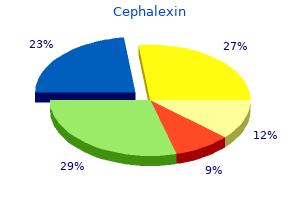
Order generic cephalexin from india. Empirical Antimicrobial Therapy in Cattle Part 10: Lumpy Jaw Wooden Tongue.