
Edward (Ted) R. Melnick, MD
- Department of Emergency Medicine
- North Shore University Hospital
- Manhasset, New York
Telson with distal end rounded herbs de provence uses cheap npxl online visa, 2 pairs of terminal spines not extending beyond distal end zever herbals purchase 30caps npxl otc, series of many plumose setae along distal margin between inner pair of terminal spines herbals and surgery purchase npxl 30 caps otc. Carapace with dark longitudinal mediolateral line on both sides; with dark brown or green diffuse spots at base of rostrum (small) and near posterior margin (large); rostrum with dorsal lamina translucent herbs used for medicine purchase npxl 30caps line, ventral lamina dark in colour herbals incense cheap npxl master card, usually bluish or greyish quincy herbals buy discount npxl. Chela of second legs in males with distinct dark blue fingers, base of fingers orange to yellow. Pleocyemata: Caridea: Palaemonoidea: Palaemonidae 129 Size: Maximum total length 18. Habitat, biology, and fisheries: Occurring in fresh, brackish and sometimes salt water. This species does not seem to occur in large quantities, thus the fishery is of a local nature. Distribution: Eastern Atlantic: West Africa from the Cape Verde Islands and Senegal to south Angola. Rostrum styliform bearing dorsally a group of 7 to 11 teeth on the basal crest, and 1 isolated tooth near the tip; its ventral margin armed with 3 to 11 teeth with single row of setae between ventral teeth. Second legs slender; fingers of chela slender, almost twice as long as palm; carpus about as long as palm; merus more than twice as long as carpus. Third, fourth and fifth legs very slender; dactylus exceptionally long and slender, almost as long as propodus; fourth and fifth legs distinctly longer than third. Telson with a truncate tip bearing 2 large, movable spines, its lateral margins with 2 pairs of small spines. Habitat, biology, and fisheries: Found on sandy and muddy bottoms in estuarine as well as coastal marine waters to depths of about 50 m. Pleocyemata: Caridea: Palaemonoidea: Palaemonidae 131 Palaemon adspersus Rathke, 1837 Frequent synonyms / misidentifications: None / None. Rostrum extending beyond tip of scaphocerite; straight, with 6 or 7 (rarely 5 or 8) regularly spaced dorsal teeth of which only 1 is positioned behind the orbit; 3 ventral teeth (rarely 2, 4, or 5); tip often bifid. Antennules triramous; shorter ramus of outer antennular flagellum normally exceeding length of peduncle and fused for third of its length to longer ramus. Colour: uniformly transparent with scattered dark spots over body, without distinct lines and bands; legs transparent with yellow at joints; characteristic for this species, compared to the other species in the area, are the many dark spots on the ventral part of the rostrum; eggs dark brown to black. Habitat, biology, and fisheries: Occurring in coastal waters at depths to 10 m, seldom deeper. Commercially important along the Atlantic coast of Europe and in the Mediterranean. On the Atlantic coast of Morocco (near Mehdiya) the species is caught and used as food. Distribution: Eastern Atlantic: Baltic from southwest Finland and Sweden south to Poland, Germany and Denmark; southern Norway and British Islands south to the Atlantic coasts of France, Spain, Portugal and Morocco; Mediterranean; Black and Caspian Seas. Pleocyemata: Caridea: Palaemonoidea: Palaemonidae 133 Palaemon elegans Rathke, 1837 Frequent synonyms / misidentifications: None / None. Rostrum straight or slightly upcurved, just reaching beyond the scaphocerite; 7 to 9 regularly spaced, dorsal teeth, tip often bifid, 3 (rarely 2) teeth situated behind the orbit; 3 ventral teeth (rarely 2 or 4). Antennules triramous; shorter ramus of outer flagellum stout, about equal in length to peduncle and fused for about two-fifth of its length to longer ramus. Scaphocerite extends to proximal half of propodus of second pereiopod (maximum); distolateral tooth not exceeding lamellar portion. Fingers of chela of second legs not more than half length of palm; carpus slightly longer than merus. Habitat, biology, and fisheries: Usually inhabiting rock pools and the shallow sublittoral zone (0 to 10 m). Of commercial interest along the Atlantic coast of Europe and in the Mediterranean. Distribution: Eastern Atlantic: from western Norway, western Sweden, Denmark and the British Islands (including the North Sea) south to southwest Africa; Mediterranean; Black Sea; Caspian Sea. Milne Edwards, 1837 Frequent synonyms / misidentifications: Palaemon garciacidi Zariquiey Alvares, 1968 / None. Scaphocerite extends to distal half of carpus of second legs, occasionally little further; apical spine not exceeding lamellar portion. Fingers of second legs usually slightly shorter than palm; dactylus and carpus slender; carpus equal to or slightly longer than merus. Distribution: Eastern Atlantic: northwest Germany, British Isles and the Netherlands south to France, Spain, Portugal and the Atlantic coast of Morocco. Pleocyemata: Caridea: Palaemonoidea: Palaemonidae 137 Palaemon macrodactylus Rathbun, 1902 Frequent synonyms / misidentifications: None / None. Antennules triramous; shorter ramus of outer antennular flagellum fused for a quarter of its length to longer ramus. Scaphocerite extends to carpus of second legs; distolateral tooth not exceeding lamina. Fingers of second legs usually slightly shorter than palm; dactylus and carpus slender, palm broad; carpus equal to length of merus. Carapace with weakly developed pattern of oblique stripes on finely dotted background. Legs brownish to reddish (sometimes translucent light bluish) with a tinge of orange at articulations and a small indistinct brownish band above them. It is expected that this species will spread along the east Atlantic coast of Europe and North Africa. Introduced in the eastern Pacific (San Francisco Bay area), Australia (Northern Territories and New South Wales) and recently in the east Atlantic (Orwell Estuary, Suffolk, eastern England; northern France; Belgium; the Netherlands; Cadiz, Spain; northern Morocco). Pleocyemata: Caridea: Palaemonoidea: Palaemonidae 139 Palaemon maculatus (Thallwitz, 1892) Frequent synonyms / misidentifications: Leander maculatus Thallwitz, 1892 / Palaemon edwardsii Rathbun, 1900. Antennules triramous; shorter ramus of outer antennular flagellum fused with 12 or 13 segments to longer ramus; shorter free ramus of flagellum as long as fused part. Carpus of second pair of legs longer than merus and chela; the propodus/carpus joint reaching beyond anterior margin of antennular scale (scaphocerite). Antennules triramous; shorter ramus of outer antennular flagellum fused for 8 articles, a third of its length, to longer ramus. Carpus of second pair of legs shorter than merus and pincer; propodus/carpus joint not reaching anterior margin of scaphocerite. Colour: pale pink, the carapace ornamented with longitudinal or oblique dark lines; eyestalks, antennules, pereiopods and telson with red blotches. Habitat, biology, and fisheries: Inhabits rocky as well as muddy bottoms in shallow waters to depths of about 40 m. Spawning occurs from mid-November to the beginning of summer; most females spawn twice, each spawn comprising 1 500 to 4 500 eggs, depending on size of female. Distribution: Eastern Atlantic: Morocco to Mauritania; northward along the coasts of Europe to Denmark; Mediterranean. Pleocyemata: Caridea: Palaemonoidea: Palaemonidae 141 Palaemon xiphias Risso, 1816 Frequent synonyms / misidentifications: None / None. Rostrum long and slender, sometimes slightly upcurved, projecting beyond tip of scaphocerite; 7 or 8 dorsal teeth of which 1 or 2 are positioned behind posterior edge of orbit, second tooth from posterior distinctly more distant from first than from third, distalmost 2 teeth separated from proximal series by toothless region; 4 or 5 ventral teeth. Antennules triramous; shorter ramus of outer antennular flagellum fused for less than fifth of length to longer ramus. Scaphocerite just overreaching carpus of second legs; distolateral spine not exceeding lamina. Fingers of second legs slender, slightly longer than somewhat swollen palm; carpus equal to or slightly shorter than merus. Absent in the Black Sea, its presence in the southeastern Mediterranean needs confirmation. Pleocyemata: Caridea: Palaemonoidea: Palaemonidae 143 Palaemonetes varians (Leach, 1814) Frequent synonyms / misidentifications: None / None. Rostrum straight, apex occasionally bifid; 4 to 6 dorsal teeth (rarely 1 to 3, 7 or 8), 2 ventral teeth (rarely none, 1, 3 or 4); 1 dorsal tooth postorbital. Outer edge of stylocerite slightly convex, anterior border straight, apical spine short and stout. Colour: completely translucent, sometimes somewhat green-brown, without stripes; pereiopods translucent with yellow at joints; white or yellow chromatophores scattered on carapace, margins of abdominal pleura and ventral margin of rostrum. Of minor importance along the European Atlantic coast where it is sometimes taken as food and sometimes used as bait. Distribution: Eastern Atlantic: west Baltic and North Sea to the Atlantic coast of Morocco; Mediterranean coast of Algeria. Rostrum well developed, laterally compressed, usually denticulate, seldom extending beyond scaphocerite. First pair of pereiopods symmetrical, ending in clearly distinct pincers, broader than second pair, but not considerably enlarged. Similar families occurring in the area Barbouriidae: carapace armed with subocular tooth posterodorsal to orbital angle. Nematocarcinidae: first 2 pairs of pereiopods similar, their carpus unsegmented, the following pairs notably elongate. Palaemonidae: first pair of pereiopods with small pincers; second pair much better developed and bearing strong pincers, their carpus unsegmented. Pandalidae: first pair of pereiopods with very small or no pincers; carpus in second pair segmented. Penaeidean shrimps: pleura of second abdominal segment not overlapping those of first segment; the 3 first pairs of pereiopods ending in pincers. Carpus of second leg with 3 or more segments; lateral surface of carapace without many small spines. Trachycaris spines on lateral surface of carapace carpus 2-segmented a) second leg b) lateral view. Rostrum with well-developed ventral lamina; series of 5 to 9 small spines at the anterior margin of the carapace (. Rostrum not ventrally developed, but might be somewhat well-developed ventral developed proximally; anterior lamina of rostrum margin of carapace without series of spines on series of spines. Carapace without pterygostomian spine; dorsal margin of rostrum with 4 widely spaced teeth (. Carapace with pterygostomian spine; dorsal margin of rostrum with 18 to 20 closelyset teeth (. Movable plate present at the movable plate distal segment of the antennular peduncle (. No movable plate present on the distal segment of the antennular peduncle; epipods on at least the bases of the first two pereiopods (. Rostrum reaching distal margin of scaphocerite; branchiostegal spine present, pterygostomian spine absent (. Rostrum not overreaching distal margin of antennular peduncle; branchiostegal spine absent, pterygostomian spine present (. Pleocyemata: Caridea: Alpheoidea: Hippolytidae 149 Hippolyte coerulescens (Fabricius, 1775). Carapace with antennal and pterygostomian spines, without branchiostegal and supraorbital spines. Second pair of pereiopods slender, their carpus divided into 13 segments, the following pairs with movable spines on the merus and a short dactylus. Telson ending in a pointed tip flanked by a movable spine on either side, its lateral margins with long hair and 2 pairs of movable spines. Habitat, biology, and fisheries: Inhabits coastal and estuarine waters, to about 15 m depth on sand and mud bottoms. Separate statistics are not reported for this species, but catches are doubtless very small, the species being only of limited local interest; in Nigeria it is reported to be caught in somewhat larger quantities (several hundred tonnes annually) combined with Nematopalaemon hastatus. Pleocyemata: Caridea: Alpheoidea: Hippolytidae 151 Lysmata seticaudata (Risso, 1816) Frequent synonyms / misidentifications: None / None. Rostrum straight, curved up at tip, rather short, not reaching end of antennular peduncle; with 5 to 7 dorsal and 2 ventral teeth; 3 (seldom 2) dorsal teeth situated behind the orbit. Carapace with large antennal and small pterygostomian spines, without supraorbital and branchiostegal spines. Antennal scale (scaphocerite) almost twice as long as antennular peduncle, outer margin slightly concave, apical spine overreaching larger part of last segment. First pair of pereiopods slender, equal in size and form, chela with black fingers; fingers much shorter than palm. Second pair of pereiopods much longer and more slender than first, distal part of ischium, merus and carpus divided in segments; carpus subdivided in many small segments (about 30). Telson with 2 pairs of dorsal spines, fringe of hairs along lateral margin of telson. Colour: body with straight, red, longitudinal bands, separated by pale bands of similar width; pereiopods and other appendages reddish. Distribution: Eastern Atlantic: West coast of Europe from the Channel Islands south to southern Spain and northern Morocco and the Canary Islands; Mediterranean; Black sea. Rostrum slender and D forward directed, not or just extending beyond eyes, with 2 small distal teeth. First pereiopods unequal, right pereiopod with distinct chela, left pereiopod simple. Second pereiopods also unequal, right pereiopod much longer than left (except in Processa modica), chela small, carpus and merus multi-articulate. Colour: translucent pink, pale brown or green, with red chromatophores, sometimes with white spots.
Systematics and Zoogeography of the worldwide bathypelagic squid genus Bathyteuthis (Cephalopoda: Oegopsida) herbs de provence uses order genuine npxl on line. Volume 1: Introduction 3-1 herbals letter draft generic npxl 30caps line, molluscs herbals on express purchase npxl 30caps with amex, crustaceans herbals for cholesterol cheap 30caps npxl with amex, hagfishes herbals and surgery generic 30caps npxl with visa, sharks herbals for weight loss cheap npxl 30 caps otc, batoid fishes and chimaeras. Dactylus of tentacular clubs with quadriserial sucker but the carpal region is greatly expanded and carries numerous small suckers in many series. Light organs known only on eyes, where a single ventral light organ may be present. Habitat, biology, and fisheries: Little is known about the biology of brachioteuthid squids although aggregations near the ocean floor, at depth of about 800 m, have been observed from submersibles. Remarks: While only 2 genera are presently recognized in this family, many species exist, most of which are not described. Similar families occurring in the area Ommastrephidae and Loliginidae: Ommastrephidae has T-shaped funnel locking apparatus; in Loliginidae the eye lens is covered by a cornea; neither of those families have numerous series of suckers in the carpal region of the tentacular clubs, a character shared with the Architeuthidae and the Neoteuthidae; in Neoteuthidae, the posterior edges of the fins are convex whereas in architeuthids the digestive gland abuts the cephalic cartilage. Mantle very narrow, widening slightly at anterior opening and tapering abruptly in front of fin; fin length and width about 50% mantle length; fin width to length ratio about 0. Mantle cylindrical, not tapering abruptly in front of fin; fin transversely rhomboidal; fin less than 50% mantle length; fin width to length ratio usually 1. Preliminary description of two new species of Cephalopods (Cephalopoda: Brachioteuthidae) from South Atlantic and Antarctic waters. In situ observations on Brachioteuthis beanii Verrill: paired behavior, probably mating (Cephalopoda, Oegopsida). Brachioteuthidae 459 Brachioteuthis picta Chun, 1910 Frequent synonyms / misidentifications: None / None. Tentacular clubs expanded, parts of the club not clearly differentiated, with at least 50 minute suckers in the carpal region that extends proximally along the club. Primarily occurring in depths of 50 to 200 m but its depth range extends from 0 to 952 m, 700 to 952 m by day; the juveniles are epipelagic; 46 to 370 km from shore off Namibia. Spermatophores are transferred to the buccal membrane of the female and spawning is in the water column. Remarks: Chun (1910) gives the type locality as Valdivia, but this is apparently an error. Habitat, biology, and fisheries: Epi mesopelagic species, adults also in bathypelagic habitat. The spawning season appears to be much extended; hence recently hatched individuals are found throughout the year. Paralarvae have long necks, planktonic and experiencing relatively deep transformations during growth. Mediterranean sea; In the Atlantic from southern Norwegian Sea and Iceland to extreme southern Atlantic, not in tropical western Atlantic, nor Gulf of Mexico and Caribbean; Indian Ocean, except Arabian Sea and North Bay of Bengal; central and southern Pacific; circumglobal in southern Ocean. Funnel-locking apparatus oval, generally Dwith 1 or 2 knobs (tragus and antitragus) directed toward the centre of the concavity. Club very elongate and divided into 2 or 3 portions by symmetrical protective membranes, except in Planctoteuthis. Very distinctive paralarvae, known as the Doratopsis stage, with an elongate neck and brachial pillar. The presence of a Doratopsis paralarva is the only character that is unique to the family. Numerous chambers in the arms, head and mantle filled with a light-weight fluid, ammonium chloride, which provides near-neutral buoyancy for the squids. Similar families occurring in the area Mastigoteuthidae: tentacles have very numerous minute suckers in more than 6 series; necks are not elongate. Funnel locks to mantle with cartilaginous apparatus; tentacle clubs with suckers 2a. With a compact club not divided into proximal and distal portions by protective membranes; arms subequal in length in adults (ventral arms much longer in young) 2b. Club very elongate and divided into 2 or 3 portions by protective membranes; ventral arms greatly elongate and thickened *Planctoteuthis. The species of this genus present in the area are compared in the following table (from Young et al. Remarks: Considerable morphological differences exist among genera of this family. Species of Planctoteuthis are usually rather small and very fragile deepsea squids, but, unlike other chiroteuthids, the subadult retains the peculiar doratopsid paralarval tentacular club. Annotated type catalogue of the Cephalopoda (Mollusca) in the Museum fur Naturkunde, Humboldt University of Berlin. Chiroteuthis veranyi from the Atlantic sector of the Southern Ocean (Cephalopoda: Chiroteuthidae). A review of the Valbyteuthidae and an evaluation of its relationship with the Chiroteuthidae. Diagnostic characters: Head and arms well developed; mantle relatively small and calyx-like in form; round terminal fins. Club sucker stalks in 2 distinct parts; stalks of lateral sucker series about twice as long as those of medial sucker series. Found in the bathyal and in the mesopelagic and upper bathypelagic zones above the continental slope from the surface to depths of 2 130 m by day and 100 to 600 m at night. Known predators are Dissostichus eleginoides (Patagonian toothfish), Prionace glauca (blue shark), Thunnus alalunga (albacore), Diomedea chrysostoma (grey-headed albatross), Diomedia exulans (wandering albatross), dorsal view Diomedea melanophrys (black-browed albatross), Globicephala malaena (pilot whale), Globicephala melas (long-finned pilot whale), Hyperoodon planifrons (bottlenose whale), Mirounga leonina (southern elephant seal), Stenella coeruleoalba (striped dolphin) and Physeter catodon (sperm whales). Widely distributed in the Atlantic: from Azores to Namibia; from the Reykjanes Ridge to South Georgia. Indian and Pacific Oceans from Tierra del Fuego to South Africa, Peru and Chile; northeastern Pacific to Oregon. Chiroteuthidae 465 Grimalditeuthis bonplandi (Verany, 1839) Frequent synonyms / misidentifications: Doratopsis sagitta Chun, 1908 / None. Arms approximately subequal in length, gelatinous; sucker base with 3 conical papillae. A very characteristic pattern of chromatophores on the head: a line of chromatophores passes across the ventral surface of the head between the anterior ends of eyes; another line runs along the neck from each olfactory papilla anteriorly to each eye, then anterior to each eye along the brachial pillar, terminating at the base of the arms. Habitat, biology, and fisheries: this species is infrequently captured but seems to have a worldwide distribution in tropical to temperate seas, inhabiting in the mesopelagic and bathypelagic zones from the surface to 2 500 m by day and 200 to 1 500 m at night. Known predators are Alepisaurus ferox (longnose lancetfish) and Xiphias gladius (swordfish). Tentacular club small, compact, with low protective membranes along both boarders, and not divided into proximal and distal regions by pro tective membranes and with a keel. Funnel locking apparatus oval with posterior bump (antitragus) double and with the lobes nearly equal. Habitat, biology, and fisheries: Occurring at depths of 0 to 2 330 m by day, 50 to 1 250 m at night. Among the deepest living of all pelagic squid, there is a ventral view dorsal view suggestion of ontogenetic vertical (illustrations from Guerra, 1992) spreading but no indication of diel vertical migration. Distribution: From Madeira to Guinea Bissau in the Atlantic; eastern Pacific from California to Chile; Hawaii and eastern Polynesian Islands. Remarks: the type species, Planctoteuthis danae, was originally placed in a new genus, Valbyteuthis, within a new family, Valbyteuthidae, by Joubin (1931). Roper and Young (1967) placed Valbyteuthis in the family Chiroteuthidae noting the similarity of Valbyteuthis paralarvae to those of Chiroteuthis. Young (1991) recognized that some paralarvae of Valbyteuthis had been previously descibed by Pfeffer (1912) as members of his new genus, Planctoteuthis, within the Chiroteuthidae. Chiroteuthidae 467 Planctoteuthis exophthalmica (Chun, 1908) Frequent synonyms / misidentifications: Doratopsis exophthalmica Chun, 1908; Chiroteuthis (Planktoteuthis) exophthalmica (Chun, 1908) / None. Funnel locking apparatus oval with posterior bump (antitragus) Light organs absent. Tentacular club small, compact; with low protective membranes along both boarders, and not divided into proximal and distal regions by protective membranes. Funnel-locking apparatus oval with posterior bump (antitragus) single or slight double, low and broad. Habitat, biology, and fisheries: this species is known for certain only from the original description, tentacular which was based on a 16 mm mantle length club specimen and the head of another, from the same haul, estimated to have a mantle length of 20 mm. Fins Dextend nearly the full length of the mantle in adults; they attach to lateral walls of mantle and are comprised of slender muscle bundles connected by membranes, producing a comb-like appearance. Occurring from the surface to 3 000 m; most abundant at depths from 500 to 1 000 m during the day and migrating into near-surface waters at night. Similar families occurring in the area None, no other family has comb-like muscle bundles in fins. No light organ on neither eyes nor viscera ventral view Remarks: this family, monotypic, is in need of revision, and a number of undescribed species are included in the only recognized genus. Along with Chtenopteryx sicula 2 other species are generally recognized at present: C. Chtenopteryx Appellof, 1890 (Mollusca, Cephalopoda): proposed confirmation as the correct original spelling. Distribution: Tropical eastern central Atlantic from Canary Islands to the Equator. Known predators include Beryx spendens (splendid alfonsino), Chauliodus (viperfish), Synaphobranchus, (deep-sea eel), Thunnus alalunga (albacore), Xiphias gladius (swordfish) and a Mediterranean dolphin. Digestive gland generally spindle-shaped and situated well posterior to cephalic cartilage. Habitat, biology, and fisheries: Cranch squids occur from the surface to approximately 2 000 m. Several species have been observed in deep water from submersibles to exhibit a peculiar posture (cockatoo posture) with the arms and tentacles folded back over the head. These can involve changing eye shape and position, changing fin shape, increased pigmentation and development of light organs on arm tips, various modifications of arm structure and, apparently, loss of tentacles. These changes have led to many developmental stages being named as separate species or genera. Most species occupy progressively deeper waters as they grow larger (ontogenetic descent). Ventral surface of mantle wit 1 or 2 cartilaginous strips extending posteriorly from anterior apex of funnel-mantle fusions; funnel fused to head laterally; eyes with 4 or more small, rond to oval photophores 1b. Ventral surface of mantle without cartilaginous strips extending posteriorly from anterior apex of funnel-mantle fusions; funnel free from head laterally; eyes with 1 usually large photophore, or 2 or 3 markedly dissimilar-sized photophores with the largest usually crescent-shaped 2a. Ventral surface of mantle with 2 cartilaginous strips in inverted V-shaped pattern extending posteriorly from anterior apex of funnel-mantle fusions; funnel valve present; dorsal pad of funnel organ with 3 longitudinal, triangular flaps; gladius with short conus 2b. Ventral surface of mantle with 1 cartilaginous strip extending posteriorly from anterior apex of funnel-mantle fusions; funnel valve absent; dorsal pad of funnel organ with 3 to 7 narrow papillae; gladius with long slender conus 474 Cephalopods 3a. Fins small, paddle-shaped, subterminal; eyes with 1, usually large, photophore 4b. Fins not paddle-shaped, may be short to long, round to lanceola-shaped, terminal or terminal lateral; eyes with 1 large and 1 or 2 small photophores 5a. Fins fused distally, inset on short rostrum of gladius which projects dorsally free of end of mantle; eyes small to medium 5b. Fins widely separated, insert on lateral expanded ends of transverse extensions of posterior end of gladius; eyes proportionally large to huge 6a. Gladial conus short, broad to narrow; fins short (<25% mantle length), oval to round; digestive gland long, narrow, spindle-shaped 6b. Gladial conus medium to long, narrow, or needle-like to filiform; fins medium to long (30 to 60% mantle length), narrow, lanceolate to ovate; digestive gland stout, spindle-shaped or rounded 7a. Posterior fin insertions do not extend to tip of gladius; no tubercles present on funnel-mantle fusion cartilages; dorsal pad of funnel organ with large, triangular lobe on each lateral arm; eyes with small, round, anterior photophore indented into median anterior margin of large, round, posterior photophore 7b. Posterior fin insertions extend to tip of gladius; 2 small tubercles present at anterior end of funnel-mantle fusion cartilages; dorsal pad of funnel organ with large, spatulate papilla on each lateral arm; eyes with small, crescent-shaped, anterior photophore lying closely within concavity of large, crescent-shaped, posterior photophore 8a. Arms with hooded hoks on midportion; fins stout, ovate (nearly round in combined outline in juvenile), do not become attenuate posteriorly 1/ this genus has no species represented in the area Cranchiidae 475 11a. Funnel valve present; dorsal pad of funnel organ with triangular flap on each lateral arm; eyes with 2 photophores (large, roughly crescent-shaped posterior photophore, and within its concavity, smaller, roughly elongate S-shaped anterior photophore); carpal suckers in 2 series on tentacular stalk 11b. Funnel valve absent; dorsal pad of funnel organ with long, spatulate papilla on each lateral arm; eyes with 3 photophores (large, crescent-shaped posterior photophore, and within its concavity, a smaller, crescent-shaped anterior photophore and a third small, oval photophore); carpal suckers in 4 series set in zigzag pattern on tentacular stalk 12a.
For individuals incontinent of urine and/or bowel identify and treat the cause when pos sible worldwide herbals purchase npxl with a mastercard. Implement a structured perineal skin care program for individuals incontinent of urine and/or feces jeevan herbals purchase npxl toronto. Cleanse the skin after each incontinence episode particularly if feces are present wiseways herbals buy npxl 30 caps overnight delivery. Use of emollients herbals that increase bleeding buy generic npxl on line, barrier products and proper incontinence containment devices that wick and hold moisture away from the skin are important aspects of a perineal skin program herbs meaning buy cheap npxl on line. Screen and assess the nutritional status of every individual at risk of pressure ulcers in each health care setting herbs mac and cheese purchase npxl 30caps free shipping. Individuals at risk of pressure ulcer development may also be at risk of under nutrition, and so should be screened for nutritional status. Use a valid, reliable and practical tool for nutritional screening that is quick and easy to use and acceptable to both the individual and health care worker. Establish a nutritional screening policy in place in all health care settings, along with rec ommended frequency of screening for implementation. Refer each individual with nutritional risk and pressure ulcer risk to a registered dieti tian and also, if needed, to a multidisciplinary nutritional team that includes a registered dietitian, a nurse specializing in nutrition, a physician, a speech and language therapist, an occupational therapist, and when necessary a dentist. Provide nutritional support to each individual with nutritional risk and pressure ulcer risk, following the nutritional cycle. Follow relevant and evidence based guidelines on enteral nutrition and hydration for individuals at risk of pressure ulcers, who show nutritional risks or nutritional problems. Ofer each individual with nutritional risk and pressure ulcer risk a minimum of 30 35 kcal per kg body weight per day, with 1. Ofer high-protein mixed oral nutritional supplements and/or tube feeding, preferably in addition to the usual diet, to individuals with nutritional risk and pressure ulcer risk because of acute or chronic diseases, or following a surgical intervention. Oral nutritional supplements are of value because many pressure-ulcer-prone patients often cannot meet their nutritional requirements via normal oral food intake. Moreover, oral nutritional supplementation seems to be associated with a signifcant reduction in pressure ulcer development, compared to routine care. Repositioning should be undertaken to reduce the duration and magnitude of pres sure over vulnerable areas of the body. Use a friction reducing slider or mechanical lifting device if assistance is required for moving. Do not position the individual directly onto medical devices, such as tubes or drain age systems. Avoid positioning the individual on bony prominences with existing non-blanchable erythema. Frequency of repositioning will be infuenced by variables concerning the individual (Strength of Evidence = C) and the support surface in use. An individualized turning schedule should be developed based on the above factors. If the individual is not responding as expected to the repositioning regime, reconsider the frequency and method of repositioning. If changes in skin conditions should occur, the repositioning plan needs to be reevaluated. Use devices to enable the individual to assist or independently position, lift and transfer. If it is necessary to raise the head of bed, limit it to no greater than 30 degrees (Diagram B) to minimize the risk of sliding and shear forces. Individuals positioned in the lateral side lying position should be positioned at 30 degrees of side lying. Individuals should be positioned in a wheel chair or other suitable chair for meals and activities. Repositioning for the Prevention of Pressure Ulcers 32 Repositioning the Seated Individual 32. Position the individual to maximize his/her functional abilities while minimizing the efects of pressure and shear. Armrests should be adjusted to the appropriate height to provide support to the upper extremities. Select a posture that optimizes the pressure redistribution and reduces shear exerted on the skin and soft tissue (Strength of Evidence = C). Limit the amount of time that an individual spends seated in the chair without some form of pressure redistribution. When an individual is seated correctly in a chair, the weight of the body still causes signifcant exposure to pressure over bony prominences. As the loaded area in such cases is relatively small, the pressure has the potential to remain high. Therefore, without adequate pressure redistribution, a pressure ulcer can develop very quickly. Individuals who are able to reposition themselves should be taught to reposition frequently, preferably every 15 minutes by using methods such as full or partial push ups, forward lean or side to side lean. Dynamic tilt or tilt used in combination with other dynamic wheelchair functions (such as recline) may be indicated for individuals who are unable to independently maintain or change their position. It has been shown that at least 30 degrees of tilt is required for substantive pressure reduction at the ischial tuberosities and the sacrum. This would include (but is not limited to) individuals with abnormal muscle tone, sensory impairment, and musculoskeletal deformities. Common observations that indicate an individual is not tolerating standard positioning include forward leaning, leaning to the side, sliding forward on the seat, feet falling of the footrests, redness on the skin (suggesting decreased tissue tolerance), and reports of discomfort. Record repositioning regimes, specifying frequency and positions adopted, and include an evaluation of the outcome of the repositioning regime. Education about the role of repositioning in pressure ulcer prevention should be ofered to all persons involved in the care of individuals at risk of pressure ulcer development, includ ing the individual and signifcant others (where possible). Training in the correct methods of repositioning and use of equipment should be of fered to all persons involved in the care of individuals at risk of pressure ulcer develop ment, including the individual and signifcant others (where possible and appropri ate). Do not base the selection of a support surface solely on the perceived level of risk for pressure ulcer development or the category/stage of any existing pressure ulcers. Ensure that the support surface is working and that the client is not bottoming out by palpating support surface areas that correlate to weight bearing bony prominences. Use higher-specifcation foam mattresses rather than standard hospital foam mat tresses for all individuals assessed as being at risk for pressure ulcer development. There is no evidence of the superiority of one higher-specifcation foam mattress over alternative higher-specifcation foam mattresses. Use an active support surface (overlay or mattress replacement) (see glossary) for individuals at higher risk of pressure ulcer development where frequent manual repositioning is not possible. Alternating-pressure active support overlays and replacement mattresses have a similar efcacy in terms of pressure ulcer incidence. Support surface selection requires consideration of the weight of the bed, the structure of the home, the width of doors, the location of power outlets, the availability of uninterrupted electrical power, and the ability to promote ventilation of heat from the motor. This should be considered in daily fuid intake and output (Australian Wound Management Association 2001). Ensure that the heels do not contact the surface of the bed for individuals at high risk of developing heel pressure ulcers. Heels can be ofoaded by placing a pillow under the calves so that heels are not touching the surface. Flex the knee slightly to avoid popliteal vein compression and increased risk of deep vein thrombosis. If pillows are not efective in ofoading heels, heel-protection devices can be con sidered. They should elevate the heel completely (ofoad them) in such a way as to distribute the weight of the leg along the calf without putting pressure on the achil les tendon. It is important to consult a clinician with expertise in using heel ofoading devices as inappropriate selection or application can put the individual at risk of developing pressure ulcers on other areas of the foot or leg. Check device placement more frequently in individuals with neuropathy, periph eral arterial disease, lower-extremity edema, or who are likely to develop edema. Use a pressure-redistributing seat cushion for individuals sitting in a chair whose mobility is reduced and who are thus at risk of pressure ulcer development. No one commercially available pressure redistributing cushion has been found to signifcantly out-perform the others in redistributing pressure (Garber et al. The seating components need to address a number of performance goals in additions to pressure redistribution. Assigned wheelchairs and seating components, including cushions, are not to be interchanged between individuals. Consideration of a pressure-redistributing surface should also be given to other sitting surfaces. Do not use synthetic sheepskin pads, cutout, ring, or donut-type devices or water-flled gloves. Water flled gloves are inefective for pressure redistribution as the heels easily move of of the glove. The recommendations that follow address the unique needs of these special populations in relation to pressure redistribution, shear reduction, and microclimate control. Refne risk assessment of individuals undergoing surgery by examining other factors that are likely to occur and will increase risk of pressure ulcer development, including: a) Length of the operation and duration of time where repositioning cannot occur b) Increased hypotensive episodes intraoperatively c) Low core temperature during surgery d) Reduced mobility on day one postoperatively (Strength of Evidence = C) 42. Use a pressure-redistributing mattress on the operating table for all individuals identifed as being at risk of pressure ulcer development. Position the patient in such a way as to reduce the risk of pressure ulcer development dur ing surgery. Elevate the heels completely (ofoad them) in such a way as to distribute the weight of the leg along the calf without putting all the pressure on the achilles tendon. Position the individual in a diferent posture preoperatively, as well as postopera tively, than the posture adopted during surgery. Consider the need to change support surfaces for individuals who cannot be turned for medical reasons such as spinal instability, hemodynamic instability and increased intracra nial pressure. Consider slow, gradual turns allowing sufcient time for stabilization of hemodynamic and oxygenation status. However, turning the individual more slowly or in small increments that allow adequate time for stabilization of vital signs should be considered when possible. Consider more frequent small shifts in position to allow some reperfusion in individuals who cannot tolerate frequent major shifts in body position. Small shifts do not replace support-surface changes when needed or turning (major shifts in body position) when pos sible. Secure the individual with bolster pads (provided by the manufacturer) to prevent sacral shearing when lateral-rotation features are selected for individuals without pressure ulcers. Discontinue lateral rotation at the frst sign of tissue damage, and re-evaluate the individual and the support surface. Confrm that the width of the bariatric individual does not reach the side rails of the bed when the individual is turned side-to-side. Consider using features that provide air fow over the surface of the skin to facili tate fuid evaporation if the skin is excessively moist or hot. Provide bariatric walkers, overhead trapezes on beds, and other devices to support contin ued mobility and independence. Pressure ulcers develop over bony prominences, but may also result from tissue pressure across the buttocks and other areas of high adipose tissue concentration. Use pillows or other positioning devices to ofoad pannus or other large skin folds and prevent skin-on-skin pressure and moisture trapping.
Loss of dorsiflexion may nations by first asking the patient to move a joint actively be due to contracture of posterior structures ridgecrest herbals anxiety free cheap 30caps npxl, such as the and then manipulating it passively when indicated herbals on demand review order npxl without a prescription. The Achilles tendon complex; loss of flexibility in the ankle gross functional tests described under the Muscle Testing syndesmosis; or impingement of anterior soft tissue or section-heel walking herbal order 30caps npxl visa, toe walking lotus herbals 3 in 1 cheap generic npxl uk, and walking on the osteophytes himalaya herbals india buy npxl on line. An ankle that cannot even dorsiflex to a neu medial and lateral borders of the feet-provide a useful tral position is said to be in equinus herbs to grow cheap npxl 30 caps with visa. For more If the Achilles tendon complex appears to be responsi detailed information, each joint needs to be isolated ble for limiting dorsiflexion, further testing may allow the individually. The principal motions of the ankle joint positions: with the knee fully extended and with the knee are dorsiflexion and plantar flexion. Extending the knee tightens the gastrocnemius motions are usually assessed with the patient in the sit portion of the Achilles complex because it originates above ting position. Flexing the knee allows the gastrocnemius to is asked to pull the foot up toward the leg as far as relax. To compare passive dorsiflexion, mius is present, less passive dorsiflexion is present with the the examiner grips the hindfoot in neutral, inverts the knee fully extended than when it is flexed. If contracture of forefoot, and passively dorsiflexes the ankle to the maxi the soleus is the limiting factor, the position of the knee mal degree possible (. Thus, any situation that causes the syndesmosis to cause such a syndrome to develop. If anterior impinge scar or to ossify in a contracted configuration also limits ment is present, passively forcing the ankle into maximal dorsiflexion. This situation commonly arises following dorsiflexion with a quick sharp movement may elicit pain sprain or fracture, especially if the ankle has been immo localized at the site of the soft tissue impingement (see bilized in a plantar flexed position. Anterior ankle impingement may also be due Finally, impingement of tissue anteriorly may also to the accretion of osteophytes on the anterior rim of the limit dorsiflexion. Such osteophytes anterior soft tissues, such as fragments of damaged liga are common among athletes in jumping sports, such as ments or tongues of inflamed synovium, may occasionally volleyball and basketball, and in ballet dancers. To assess active plantar flexion, the prominent processus trigonum or os trigonum. The patient is asked to point the fool downward as far as pos processus trigonum is a normal bony prominence of the sible (. Passive plantar flexion may be posterior talus, and the os trigonum is a small ossicle that assessed by again grasping the hindfoot, inverting the occurs at the same location. If posterior impingement is forefoot, then plantar flexing the ankle as far as possible suspected, the examiner may test for it by quickly and (. Active inver tracture may be a nonspecific sequela of a major ankle sion and eversion may be roughly assessed in the seated injury such as a fracture. Forefoot abduction and amount of motion is assessed by estimating the angle adduction are usually evaluated by passively stabilizing between a line bisecting the heel and another line bisect the calcaneus in neutral position with one hand and ing the posterior leg (. Severe tarsal joints are responsible for the small amount of restriction or absence of subtalar motion in a child or motion that exists. Clinically, it is sufficient to document that an older individual, it is the common sequela of hindfoot motion is present because it is very difficult to measure fractures, particularly those involving the calcaneus. The other hand joints are capable of extension, or dorsiflexion, and flex then grasps the great toe and passively dorsiflexes, then ion, or plantar flexion. Passive extension of two joints is normally tested simultaneously by asking the the normal first metatarsophalangeal joint averages patient to extend the great toe as far as possible (. Restriction of motion the individual movements of these two joints may in this joint is commonly called hallux rigidus. Loss of extension is par toe, active motion at these joints is usually assessed ticularly disabling because it interferes with the heel rise simultaneously by asking the patient to maximally necessary for normal gait. It is not normally possible for an indi great toe is measured in a similar manner. In this case, the vidual to isolate movement of any one of these toes examiner grasps the proximal phalanx with one hand and from the others. These motions vary considerably among Because the lesser toes each have three phalanges, they individuals. In the smaller toes, the middle phalanx is also each have three joints: the metatarsophalangeal quite short, making it difficult to distinguish distal inter joint, the proximal interphalangeal joint, and the dis phalangeal joint motion from proximal interphalangeal Figure 7-44. Passive motion of the distal interphalangeal joints of the lesser toes (third toe). As in the great toe, loss of motion in the considered abduction and motion toward the second toe, metatarsophalangeal joints has the greatest functional adduction. Loss of motion in the interphalangeal joints asking the patient to spread the toes (. This is is common following fracture or in the presence of ham not normally measured in degrees but merely by noting mer toe, claw toe, or mallet toe deformities. Similarly, active significance of the associated contractures of these joints adduction may be documented by asking the patient to is their tendency to cause friction against the adjacent squeeze the toes together. As in the hand, abduction is considered motion Palpation is usually performed with the patient seated away from the midline of the foot, rather than the with the lower part of the leg dangling comfortably off midline of the body. For comfort, relation to the second toe, which is considered the mid the examiner is usually seated on a low stool. It forms the anterior portion of the syndesmotic complex, which stabilizes the ankle mortise. Tenderness over the anterior-inferior tibiofibular liga ment may be associated with a syndesmosis sprain or a Maisonneuve fracture. In the more severe injuries, the tenderness continues up the leg owing to injury of the interosseous membrane. The tibialis anterior tendon may be easily identified by asking the patient to actively dorsiflex the ankle. At the level of the ankle joint, the tendon is closer to the medial malleolus than to the lateral malleolus, but the muscle itself lies lateral to the tibial crest in the anterior com partment. Peritendinitis, or inflammation of the tissue around the tibialis anterior tendon, occasionally occurs where the more proximal portion of the tendon courses over the anterior surface of the tibia (see. If the examiner lightly palpates the tendon at this point while the patient alternately dorsitlexes and plantar flexes the ankle, soft tissue crepitus surrounding the tendon is often appreciated. Acute or chronic degenerative ruptures of the tibialis anterior tendon occasionally occur. In such cases, active dorsiflexion of the ankle is accompanied by invol resistance and stability while palpating with the other untary eversion of the foot due to overpull of the exten hand. A common mistake is to use too little or too much sor hallucis longus and the extensor digitorum force when palpating. Although stress fractures more com pressed at the point where it pierces to the deep fascia of monly occur on the posteromedial aspect ot the tibia, the ankle to supply sensation to the dorsum of the foot. Laceration of the superficial per and sometimes a small visible bump at this location. The extensor hallucis longus tendon crosses the exercise-induced anterior compartment syndrome. In such artery at about the level of the ankle, so that in the ankle individuals, the anterior compartment muscles seem and the foot, the artery is located immediately lateral to normal at rest but tender and abnormally firm if the the extensor hallucis longus tendon. The best place for examiner palpates them immediately after the patient has palpating the dorsalis pedis pulse is on the dorsum of the exercised. The physical findings of the exercise-induced foot at a point just lateral to the extensor hallucis longus anterior compartment syndrome are transient and subtle tendon and just proximal to the prominence of the compared with those of the classic acute compartment metatarsal-cuneiform joints (. The tendons of the extensor digitorum longus and the adjacent peroneus tertius lie just lateral to the dorsalis pedis artery. Just distal to the ankle joint, these tendons are restrained by the interior extensor retinaculum. These tendons are usually readily visible and palpable, especially when the patient is actively dorsiflexing the toes (see. Although the tendons themselves are rarely involved in a pathologic process other than laceration, pres sure from shoes laced excessively tight can cause a tenosyn ovitis on the dorsum of the foot surrounding the extensor digitorum longus tendons. This condition may cause diffuse tenderness and mild swelling around the extensor digitorum longus tendons in the vicinity of the tarsometatarsal joint. Maximal passive plantar flexion of the ankle exposes the anterolateral aspect of the talar dome to palpation (. With this maneuver, the anterolateral por tion of the talar dome becomes palpable and often visible between the lateral malleolus and the extensor digitorum longus tendon (. Tenderness of the talar dome may reflect osteochondritis dissecans or a tran extensor retinaculum over the dorsum of the foot. Trauma, tight shoes, and space-occupying lesions are the While maintaining maximal plantar flexion of the most common causes of this condition, known as ante ankle, the examiner may palpate the tarsal navicular and rior tarsal tunnel syndrome. To check for anterior tarsal intervening talonavicular joint just distal to the ankle. In the advanced degeneration of the talonavicular joint is pres presence of an anterior tarsal tunnel syndrome, such per ent. Tenderness of the body of the navicular in an athlete cussion usually generates a burning pain that radiates to should cause the examiner to suspect a navicular stress Figure 7-51. Isolated tenderness of anterior tendon distally to its insertion leads the exam the fifth metatarsophalangeal joint may be associated iner to the medial cuneiform. Tenderness in these bones is usually mity is not always tender, active bursitis may occur over attributable either to posttraumatic arthrosis or to the prominence in extreme cases. Prominent osteophytes may be the spaces between the metatarsal heads should be identified in these circumstances. The examiner may either Further distally, the examiner palpates the articula compress the tissue between the thumb and the index fin tions of the five metatarsals with the cuneiforms and the ger of one hand or support the forefoot with one hand cuboid. These articulations are collectively called the tar while compressing with the index finger of the other (. The subtler forms of these injuries are diffi If the neuroma is very large, the examiner may actually be cult to diagnose radiographically. For exam the individual metatarsals should be identified and ple, a neuroma in the interspace between the third and palpated for tenderness. Localized tenderness suggests the fourth metatarsal heads may be associated with sensory possibility of a stress fracture. These stress fractures occur changes of the lateral side of the third toe and the medial most commonly in the shafts of the second and the third side of the fourth toe. Stress fractures of the spread so that heloma molle, soft interdigital corns, may fifth metatarsal tend to occur in the proximal metaphysis about 2 cm distal to the palpable base of the metatarsal. Such stress fractures are often called Jones fractures, although Jones fractures may also occur owing to acute trauma as well as recurrent stress. The first metatarsopha langeal joint may be tender medially in the presence of a bunion or hallux valgus deformity, although in such cases the associated deformity should be obvious. Osteo arthritis of the first metatarsophalangeal joint may pro duce a subtler finding of palpable osteophytes on the dorsum of the joint without associated angular defor mity. Extreme tenderness, especially when associ ated with swelling, warmth, and erythema around the joint, is more suggestive of acute gout or septic arthritis. The four lesser metatarsophalangeal joints should also be palpated for tenderness. Synovitis can occur owing to active rheumatoid arthritis, or it can be the result of pressure overload in a foot with a shortened, hypermobile first metatarsal that increases pressure distribution to the lesser metatarsophalangeal Figure 7-52. Below the peroneal tendons, the lateral aspect of the cal Ingrown toenails should be palpated for tenderness and caneus is subcutaneous and easily palpated. In the pres of the calcaneus in an athlete suggests the possibility of a ence of such an infection, palpating the nail border may calcaneal stress fracture. On the lateral aspect of the foot and the the sinus tarsi is a space between the lateral talus ankle, the lateral malleolus provides orientation. The accompanied by localized edema or ecchymosis, suggests sinus tarsi may be palpated as a depression immediately the possibility of a fracture (see. Slight inversion a displaced fracture seen acutely, the examiner may actu of the heel accentuates the space, allowing deeper palpa ally be able to palpate a step-off at the fracture site or feel tion to the lateral talar neck. In the presence of often indicates injury or arthritis involving the posterior an unstable fracture, the examiner may note crepitus facet of the subtalar joint. Distal to the sinus tarsi, the examiner can palpate the the lateral malleolus is also the principal landmark bony prominence of the anterior process of the for palpating the lateral ankle ligaments. This process may fracture owing to a twisting anterior flare of the lateral malleolus with the talar neck, injury. Detecting tenderness over the anterior process of the anterior talofibular ligament is the most common the calcaneus is important because such fractures are ankle ligament to be injured (see. Although the margins of the joint and ecchymosis, is clinical evidence of a sprain of the lig may be difficult to palpate, alternate abduction and ament. The functional integrity of the anterior talofibular adduction of the forefoot may allow the examiner to ligament is assessed with the anterior drawer test, identify it. Tenderness of this articulation may be due to described in the Manipulation section.
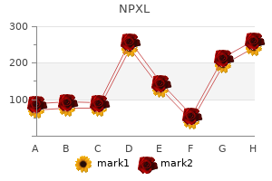
Buy npxl 30 caps with mastercard. Karta Purkh Singh Khalsa: Ayurvedic Herbalism.